Friday, April 4, 2025
Home Blog Page 15

T4 Ligation-PDF

Overview

Standard DNA ligation using NEB T4 ligase to form a vector.

Materials

For a 10 μL ligation reaction:

  • 3 μL sterile ddH2O
  • 1 μL 10X ligation buffer
  • 5 μL ~100nM linker dsDNA (either hybridization product or digested, purified PCR product)
  • 0.5 μL ~10nM backbone dsDNA (gel extracted digestion product from donor plasmid)
  • 0.5 μL T4 ligase

(Note: ratio of backbone to linker given here is ~1:100. This may need to be adjusted in some cases.)

Procedure

  1. Add the reaction components together in order listed above. Mix gently and spin.
  2. Include -linker control reaction (sub sterile ddH2O for linker).
  3. Incubate 16 °C overnight (8 hr minimum).
  4. Transform 2.5 μL rea(or more, if necessary, to obtain an acceptable amount of colonies) into chemically competent cells.


RNA extraction using trizol/tri-PDF


Overview

RNA extraction with TRIzol (Invitrogen product name) or the equivalent TRI (Sigma-Aldrich product name) is a common method of total RNA extraction from cells based on the research of Chomczynski P, Sacchi N. 1987 [1] and reviewed by the authors again in 2006 [2]. It takes slightly longer than column-based methods like RNAeasy b

ut it has higher capacity and can yield more RNA. Along with chaotropic lysis buffers it is generally considered the method that gives the best quality RNA.

Principle

  • guanidinium isothiocyanate (powerful protein denaturant) -> inactivation of RNases
  • acidic phenol/chloroform -> partitioning of RNA into aqueous supernatant for separation

Note: low pH is crucial since at neutral pH DNA not RNA partitions into the aqueous phase. Check the pH of old TRIZOL/TRI reagents!

Reagents

  • TRIzol or TRI reagent
If you want to make your own reagents, see here
RNA extraction using self-made guanidinium-acid-phenol reagents
  • 0.8 M sodium citrate / 1.2 M NaCl
  • isopropanol (2-propanol)
  • chloroform
  • 75% EtOH in DEPC H2O
  • RNase free water (filtered or DEPC)

Steps

cell lysis

Cell lysis only takes a few minutes per well, but tissue homogenisation can take 10-20 minutes per sample depending on how tough the tissue is.

  • (PBS wash)
  • add trizol (cell lysis)
1ml / 3.5 cm diameter well (6-well)
5ml / 75 ml bottle
  • homogenise by pipetting several times (mechanic lysis)
alternative for tubes: vortex 1 min
alternative for tissue: grind 1 g tissue in liquid nitrogen in a motar and pestle, put tissue into plastic screw-cap centrifuge tube + 15 ml TRIzol reagent, incubate samples for 5 min at room temp or 60° C (scaled up as needed)
  • (5min at RT for complete dissociation of nucleoprotein complexes)

RNA is stable in trizol which deactivates RNases. You can take a break at this point keeping the sample in trizol for a short time or freezing it for a longer one.

phase separation

15-45 min depending on number of samples and whether an additional chloroform wash is necessary

  • add chloroform (1/5 volume of trizol; e.g. 0.2ml to 1ml)
  • shake for 15 sec (Eccles protocol: do not vortex)
  • incubate 2-5 min at RT
  • spin max. 12000g, 5-15 min, 2-8°C
if centrifugation hasn’t been sufficient the DNA-containing interphase will be cloud-like and poorly compacted

If supernatant appears turbid an additional chloroform cleaning step can be inserted here.

  • transfer aqueous upper phase into new tube

Take care not to aspirate the DNA-containing white interface. This quickly happens and will lead to DNA contamination in your RNA prep.

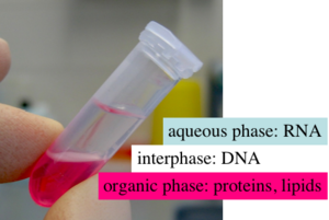
TRIZOL phases after chloroform addition
 
TOP    - colourless aqueous phase              (RNA) - 60% TRIZOL volume
MIDDLE - interphase                            (DNA)
BOTTOM - red (organic) phenol-chloroform phase (proteins & lipids)

RNA precipitation and wash

20-40 min depending on number of samples

  • add isopropanol (70% of aqueous phase or 1/2 trizol volume)
  • 0.8 M sodium citrate or 1.2 M NaCl can be added
  • (incubate 10min at RT)
  • spin max g, 10-15 min, 4ºC
  • remove supernatant
  • (alternative RNA precipitation – RNeasy from Qiagen) better than alcohol precipitation for smaller amounts of RNA (less risk of losing a miniscule nucleic acid pellet); also reduces risk of organic solvent contamination

similar kits to RNeasy: MinElute kit, or Affymetrix sample clean-up

RNA wash

15-30 min depending on number of samples

  • wash pellet 70% EtOH (add & vortex briefly)
70% ethanol prepared with RNase-free water

some prefer to wash the pellot more than once with 70% ethanol

  • spin max g, 2-10 min, 4ºC
  • air-dry pellet for 5-10 min Do not overdry the pellet or you won’t be able to redissolve it.

optional add RNase inhibitor

incubate at 55-60 C° for 10 min if hard to redissolve

  • transfer to eppendorf tube
  • spin 4° C, 5 min (to pellet undissolved material)

redissolving of RNA

  • dissolve pellet in 50-100 µl filtered or DEPC H2O (note: DEPC inhibits RT reaction)
  • alternatively, 0.5% SDS

pipetting up and down, heat to 55-60°C for 10 min

Common mistakes

  • use too little trizol; very small volumes are hard to separate and will most likely lead to contamination
  • aspirate some white interphase (DNA) when removing aqueous supernatant (RNA)
  • use phenol/chloroform of the wrong pH (has to be acidic)
  • not working under the hood (phenol is toxic , chloroform is a narcotic )

DNA Digestion-PDF

Overview

Standard DNA digest protocol using NEB restriction enzymes.

Materials

For a DNA digestion of total volume V (in μL):

  • ~0.8V DNA
  • 0.1V 10X digestion buffer (1, 2, 3, or 4; NEB)
  • 0.1V 10mg/ml BSA
  • 1 μL restriction enzyme 1
  • 1 μL restriction enzyme 2 (optional, for double digestion)

Procedure

  1. Find appropriate enzymes(s), associated buffer, reaction temperature from NEB.
  2. Calculate the total volume of the reaction by taking the DNA volume + 1(2) μL for a single (double) digestion reaction. Divide this number by 0.8 to get the total reaction volume, V.
  3. Calculate the volume of 10mg/ml BSA and 10X digestion buffer, which are both 0.1V.
  4. Add the reaction components together in order listed above. Mix gently and spin.
  5. Incubate at reaction temperature for 3 hr to overnight.

Notes

Please feel free to post comments, questions, or improvements to this protocol. Happy to have your input!

  1. List troubleshooting tips here.
  2. You can also link to FAQs/tips provided by other sources such as the manufacturer or other websites.
  3. Anecdotal observations that might be of use to others can also be posted here.

Please sign your name to your note by adding ”’*~~~~”’: to the beginning of your tip.


RNA Quality Control-PDF

Quality control of RNA

RNA Quality Control This protocol will assist you in performing RNA quality control for a variet of purposes; Microarray, real-time and Northern analysis.

Checking RNA/cRNA samples on a nanodrop Spec

  • Check concentration, A260/A280, A260/A230 on Nanodrop Spectrophotometer.
  • Maintain the A260/A280 ratio close to 2.0 for pure RNA (ratios between 1.9 and 2.1 are acceptable).
  • For Affymetrix labeling reactions; concentrations of cRNA are generally around 1000-1500ng/µl for a 40µl IVT reaction using 20µl of cDNA template.

Checking RNA/cRNA samples on an Agilent Bioanalyzer

Agilent BioAnalyzer Gel electrophoresis of the RNA is done to estimate the quality of the RNA. The bioanalyzer software produces a RIN (RNA integrity number) which gives a measure of sample quality that is not subjective. However it needs to be used with some caution and is not as useful as it sounds.

RNA Image galleries

The following are examples of typical Human RNA examined on an Agilent 2100 BioAnalyser. RNA electropherograms have different profiles depending on species, tissue and starting material quality.

  • Human RNA good quality
  • Mouse total RNA undegraded, RIN 10
  • Mouse total RNA slightly degraded, RIN 9.4
  • Arabidopsis RNA good quality
  • Arabidopsis single cell aRNA
  • Barley RNA good quality
  • Wheat RNA good quality

cRNA Image galleries

The following are examples of typical cRNA products examined on an Agilent 2100 BioAnalyser. cRNA electropherograms have different profiles depending on species and starting material quality.

  • Affymetrix One cycle Human cRNA good quality
  • Affymetrix Two cycle Human cRNA good quality
  • Affymetrix one cycle Arabidopsis cRNA good quality
  • Affymetrix one cycle Arabidopsis single cell aRNA cRNA
  • Affymetrix one cycle Barley cRNA good quality
  • Affymetrix one cycle Wheat cRNA good quality
  • Affymetrix one cycle Wheat cRNA poor quality

Affymetrix Target Hybridization-PDF

Affymetrix Eukaryotic Gene Expression Sample Processing

This protocol will allow you to perform the hybridization, washing, staining, and scanning of Affymetrix GeneChip microarrays. This protocol is a supplement to instructions provided in the Affymetrix Expression Manual.

Workflow

Materials

List reagents, supplies, and equipment necessary to perform the protocol here. For those materials which have their own OWW pages, link to that page. Alternatively, links to the suppliers’ page on that material are also appropriate.

  • Hybridisation controls: These are part of the complete One-Cycle Target Labeling and Control Reagents, Affymetrix, P/N 900493; and may be ordered separately if necessary Usually not!)
    • GeneChip Eukaryotic Hybridization Control Kit, Affymetrix, P/N 900454 (30 reactions)
    • GeneChip Eukaryotic Hybridization Control Kit, Affymetrix, P/N 900457 (150 reactions)
    • Control Oligo B2, 3 nM, Affymetrix, P/N 900301

Other reagents:

  • Water, Molecular Biology Grade, BioWhittaker Molecular Applications / Cambrex, P/N 51200
  • Bovine Serum Albumin (BSA) solution (50 mg/mL), Invitrogen Life Technologies, P/N 15561-020
  • Herring Sperm DNA, Promega Corporation, P/N D1811
  • 5M NaCl, RNase-free, DNase-free, Ambion, P/N 9760G
  • MES hydrate SigmaUltra, Sigma-Aldrich, P/N M5287
  • MES Sodium Salt, Sigma-Aldrich, P/N M5057
  • EDTA Disodium Salt, 0.5M solution (100 mL), Sigma-Aldrich, P/N E7889
  • DMSO, Sigma-Aldrich, P/N D5879
  • Surfact-Amps 20 (Tween-20), 10%, Pierce Chemical, P/N 28320
  • R-Phycoerythrin Streptavidin, Molecular Probes, P/N S-866
  • 5M NaCl, RNase-free, DNase-free, Ambion, P/N 9760G
  • PBS, pH 7.2, Invitrogen Life Technologies, P/N 20012-027
  • 20X SSPE (3M NaCl, 0.2M NaH2PO4, 0.02M EDTA), BioWhittaker Molecular Applications / Cambrex, P/N 51214
  • Goat IgG, Reagent Grade, Sigma-Aldrich, P/N I 5256
  • Anti-streptavidin antibody (goat), biotinylated, Vector Laboratories, P/N BA-0500
  • Tygon Tubing, 0.04″ inner diameter, Cole-Parmer, P/N H-06418-04
  • Tough-Spots, Label Dots, USA Scientific, P/N 9185-0000


Reagent Preparation

  • 12X MES Stock Buffer (1.22M MES, 0.89M [Na+]) 250mL
    • 16.15g of MES hydrate
    • 48.325g of MES Sodium Salt
    • 200 mL of Molecular Biology Grade water
    • Mix and when fully dissolved adjust volume to 250 mL. The pH should be between 6.5 and 6.7. Filter through a 0.2 µm filter. Do not autoclave. Store at 2°C to 8°C and shield from light. Discard solution if yellow.


  • 2X Hybridization Buffer (Final 1X concentration is 100 mM MES, 1M [Na+], 20 mM EDTA, 0.01% Tween-20) 50 mL
    • 8.3 mL of 12X MES Stock Buffer
    • 17.7 mL of 5M NaCl
    • 4.0 mL of 0.5M EDTA
    • 0.1 mL of 10% Tween-20
    • 19.9 mL of water
    • Do not autoclave. Store at 2°C to 8°C, and shield from light. Discard solution if yellow.


  • 1X Hybridization Buffer (100 mM MES, 1M [Na+], 20 mM EDTA, 0.01% Tween-20) 50 mL:
    • 25 mL of 2X Hybridization Buffer
    • 25 mL of water


  • Wash Buffer A: Non-Stringent Wash Buffer (6X SSPE, 0.01% Tween-20) 1,000 mL:
    • 300 mL of 20X SSPE
    • 1.0 mL of 10% Tween-20
    • 699 mL of water
  • Filter through a 0.2 µm filter


  • Wash Buffer B: Stringent Wash Buffer (100 mM MES, 0.1M [Na+], 0.01% Tween-20) 250 mL:
    • 20.825 mL of 12X MES Stock Buffer
    • 1.3 mL of 5M NaCl
    • 0.25 mL of 10% Tween-20
    • 227.625 mL of water
    • Filter through a 0.2 µm filter
    • Store at 2°C to 8°C and shield from light. Keep this in the bottle shielded with foil during use.


  • 2X Stain Buffer (Final 1X concentration: 100 mM MES, 1M [Na+], 0.05% Tween-20) 250 mL:
    • 41.7 mL of 12X MES Stock Buffer
    • 92.5 mL of 5M NaCl
    • 2.5 mL of 10% Tween-20
    • 113.3 mL of water
    • Filter through a 0.2 µm filter
    • Store at 2°C to 8°C and shield from light


  • 10 mg/mL Goat IgG Stock
    • Resuspend 50 mg in 5 mL of 150 mM NaCl
    • Store at 4°C
    • If a larger volume of the 10 mg/mL IgG stock is prepared, aliquot and store at -20°C until use. After the solution has been thawed it should be stored at 4°C. Avoid additional freezing and thawing.


Protocol

These protocols provide detailed steps for preparing the eukaryotic hybridization mix containing labeled target and control cRNA. After completing the procedures described in this chapter, the hybridized probe array is ready for washing, staining, and scanning, as detailed in Washing, Staining and Scanning GeneChip Arrays.

Notes before starting

  • It is imperative that frozen stocks of 20X GeneChip Eukaryotic Hybridization Controls are heated to 65°C for 5 minutes to completely resuspend the cRNA before aliquotting.
  • It is important to allow the arrays to equilibrate to room temperature completely. Specifically, if the rubber septa are not equilibrated to room temperature, they may be prone to cracking, which can lead to leaks.
  • Eukaryotic hybridisations are generally done at 45°C.
  • Streptomyces hybridisations are done at 50°C. CHECK PACKAGE INSERTS!

Setting up hyb cocktail

Please refer to the table below for the necessary amount of cRNA required for appropriate probe array format. These recipes take into account that it is necessary to make extra hybridization cocktail due to a small loss of volume (10-20 µL) during hybridization.

  • Make a master mix of hybridisation cocktail reagents and add to individual fragmented cRNAs in 1.5ml tubes.
  • If using the GeneChip IVT Labeling Kit to prepare the target, a final concentration of 10% DMSO needs to be added in the hybridization cocktail for optimal results.

Physical preparation of the array hybridisation

  • Equilibrate probe array to room temperature immediately before use.
  • Heat the hybridization cocktail to 99°C for 5 minutes in a thermomixer.
  • Wet the array by filling it through one of the septa (see below) with 1X Hybridization Buffer using a pipette and appropriate tips.

It is necessary to use two pipette tips when filling the probe array cartridge: one for filling and the second to allow venting of air from the hybridization chamber.

  • Incubate the probe array filled with 1X Hybridization Buffer at 45°C (50°C for Streptomyces) for 10 minutes with rotation at 60rpm.
  • Transfer the hybridization cocktail that has been heated at 99°C, to a 45°C (50°C for Streptomyces) thermomixer for 5 minutes.
  • Spin hybridization cocktail(s) at maximum speed in a microcentrifuge for 5 minutes to remove any insoluble material from the hybridization mixture.
  • Remove the buffer solution from the probe array cartridge.
  • Fill with appropriate volume of the clarified hybridization cocktail, avoiding any insoluble matter at the bottom of the tube. Label the array with the JobID_SampleID!
  • Place probe array into the Hybridization Oven, set to 45°C (50°C for Streptomyces). Avoid stress to the motor; load probe arrays in a balanced configuration around the axis. Rotate at 60rpm.
  • Hybridize for 16 hours. During the latter part of the 16-hour hybridization, proceed to Washing, Staining, and Scanning and prepare reagents required immediately after completion of hybridization.

Washing, Staining, and Scanning

These protocols provide detailed instructions for using the Fluidics Station 450 and GeneChip Scanner 3000 to automate the washing, staining and scanning of GeneChip expression probe arrays. After completing the procedures described in this chapter, the scanned probe array image (.dat file) is ready for analysis. After 16 hours of hybridization, remove, and keep, the hybridization cocktail from the four arrays you are washing and fill completely with Non-Stringent Wash Buffer (Wash Buffer A), as given the table at step 1 of Eukaryotic Target Hybridization. This procedure takes approximately 90 minutes to complete.


Step 1: Starting up workstation, fluidics and scanner. Turn on PC and after you have logged in start GCOS software. Turn on the fluidics station using the toggle switch on the lower left side of the machine.. Turn on the scanner (it is recommended to wait until nearly all your arrays are ready for scanning. The autoloader should allow you to walk away and the scanner does make quite a lot of noise).

Step 2: Entering Experiment Information To wash, stain, or scan a probe array, an experiment must first be registered in GCOS. The fields of information required for registering experiments in GCOS are: Sample Name: user-supplied-sample-name_JobID_SampleID_s e.g. Col1_39_7_s Sample Type Project Experiment Name: same as above without _s e.g. Col1_39_7 Probe Array Type

You should also enter the following fields if possible: Barcode – Scan in the barcodes when possible as this allows GCOS to attach a .dat file to the experiment.

The Project, Sample Name, and Experiment Name fields establish a sample hierarchy that organizes GeneChip gene expression data in GCOS. In terms of the organizational structure, the Project is at the top of the hierarchy, followed by Sample Name, and then Experiment Name.

Step 3: Preparing the Fluidics Station The Fluidics Station 450 is used to wash and stain the probe arrays. It is operated using GCOS. Setting Up the Fluidics Station Turn on the Fluidics Station if you have not already done so. Place Wash A and Wash B buffers on the fluidics station and make sure tubing is all the way inside the bottle so no air will be sucked up. Wash B is stored in the fridge and should be kept as dark as possible during use. Make sure there is enough buffer in each bottle for the number of chips you are going to run. Select Run → Fluidics from the menu bar. Or click the fluidics button on the toolbar; you will be presented with the fluidics station window (see below).

Priming the Fluidics Station Priming ensures that the lines of the fluidics station are filled with the appropriate buffers and the fluidics station is ready for running fluidics station protocols. Priming should be done: when the fluidics station is first started. when wash solutions are changed. before washing, if a shutdown has been performed. if the LCD window instructs the user to prime.

Select the Prime_450 protocol and choose All Modules, then click the Run button (this can be run on individual modules if you are only running one or two arrays.)


Step 4: Preparing the Staining Reagents Prepare the following reagents. Volumes given are sufficient for one probe array. Always prepare the stain solutions fresh, on the day of use.

Streptavidin Phycoerythrin (SAPE) should be stored in the dark at 4°C, foil-wrapped. Mix SAPE well before preparing stain solution. Do not freeze SAPE.

SAPE Solution Mix

Master Mix 2X Stain Buffer 600.0 µL 50 mg/mL BSA 48.0 µL 1 mg/mL Streptavidin Phycoerythrin (SAPE) 12.0 µL DI H20 540.0 µL

Antibody Solution Mix

Master Mix 2X Stain Buffer 300.0 µL 50 mg/mL BSA 24.0 µL 10 mg/mL Goat IgG Stock 6.0 µL 0.5 mg/mL biotinylated antibody 3.6 µL DI H20 266.4 µL

Aliquot stain solutions into tubes marked 1 & 3 (SAPE Stain) and 2 (Antibody) with 600 µL in each tube for use on the fluidics station.

Step 5: Washing and Staining the Probe Array using the Fluidics450 1. In the Fluidics Station dialog box in GCOS, select the correct experiment name from the drop-down Experiment list. (The Probe Array Type appears automatically, check that it corresponds to the array type you are using.) 2. Select the appropriate protocol from the drop-down Protocol list, to control the washing and staining of the probe array format being used: Check array package insert for correct fluidics protocol to use. 3. Choose Run in the Fluidics Station dialog box to begin the washing and staining. Follow the instructions in the LCD windows on the fluidics station modules. 4. Insert the appropriate probe array into the designated module of the fluidics station while the cartridge lever is in the down, or eject position. When finished, verify that the cartridge lever is returned to the up, or engaged, position. 5. Remove any microcentrifuge vial remaining in the sample holder of the fluidics station module(s) being used. 6. When prompted “Load Vials 1-2-3,” place the three experiment sample vials (the microcentrifuge vials) into the sample holders 1, 2, and 3 on the fluidics station. Place one vial containing 600 µL of SAPE solution in sample holder 1. Place one vial containing 600 µL of antibody solution in sample holder 2. Place one vial containing 600 µL of SAPE solution in sample holder 3. Press down on the needle lever to snap needles into position and to start the run. The Fluidics Station dialog box at the workstation terminal and the LCD window display the status of the washing and staining progress. Each run takes about 90 minutes. 7. When prompted “eject cartridge”, remove the array and check for air bubbles. If bubbles are present they need to be removed prior to scanning. Follow instructions on Removing Air Bubbles Before Scanning. 8. When prompted “remove vials”, replace the microcentrifuge vials containing stain solutions with three clean empty microcentrifuge vials. The fluidics station will now take about 20 minutes to complete the protocol and then you are ready to start the next four chips.

Remove and bubbles from the arrays. (See optional step next.)

If you do not scan the arrays immediately, store them at 4°C and in the dark until ready for scanning. Arrays can be stored for 24hours after washing and staining is complete.

When you have finished all array processing you must run the shutdown protocol with all three reagent lines in water.

Once a month you should run the Bleach cleanup protocol, detailed in the Affymetrix manual.


Step optional: Removing Air Bubbles Before Scanning. You must remove any air bubbles before scanning. The easiest way is to insert a 200 µLtip into the top septa and use another 200 µL tip to remove about half of the wash A inside the array. Still holding the pipette in the same position, remove the tip and fill with new wash A, insert it back into the lower septa and fill the array making sure you do not introduce a new bubble.

Probe Array Scan

The scanner is controlled by GCOS. The array is scanned after the wash protocols are complete. Make sure the laser is warmed up prior to scanning by turning it on at least 10 minutes before use. If probe array was stored at 4°C, it will need to warm to room temperature before scanning; a dialogue box will prompt for this when you start any scan.

Handling the GeneChip Probe Array before a Scan If necessary, before you scan the probe array, clean the glass surface with a non-abrasive towel or tissue. Do not use alcohol to clean glass. Before scanning the probe array cartridge, apply Tough-Spots™ to each of the two septa on the probe array cartridge to prevent the leaking of fluids from the cartridge during scanning.

1. On the back of the probe array cartridge, clean excess fluid from around septa. 2. Carefully apply one Tough-Spot to each of the two septa. Press to ensure that the spots remain flat. If the Tough-Spots do not apply smoothly, that is, if you observe bumps, bubbles, tears, or curled edges, do not attempt to smooth out the spot. Remove the spot and apply a new spot. The scanner uses a laser and is equipped with a safety interlock system. Defeating the interlock system may result in exposure to hazardous laser light. You must have read, and be familiar with, the operation of the scanner before attempting to scan a probe array.

Scanning the Probe Array The Scanner 3000 is fitted with an autoloader. This is cooled and must be loaded from position 1 which is outlined in red. Chips can only be loaded in the correct orientation. 1. Select Run → Scanner from the menu bar. Alternatively, click the Start Scan icon in the tool bar. The Scanner dialog box appears with a drop-down list of experiments that have not been run. 2. Verify whether the arrays are at room temperature or not. 3. Click the Start button. 4. Each array will take about 5 minutes to scan. The autoloader and GCOS should allow you to walk away as the scan is organised by the barcode on the arrays.

If you need to the scanner can be operated in manual mode with or without the autoloader. Refer to the scanner Quick Reference Card.


Affymetrix data analysis and formatting.

GCOS analysis

Data needs to be analysed in GCOS and the formatted for QC analysis and customer use.

  • Check analysis settings for the array type you are analyzing.

Scaling should be ‘All Probe Sets’ and ‘Target Signal’ should be 100.

  • Select the .cel files you want to analyse, right click and select ‘Analyze’.
  • Save the pivot data table as a tab delimited txt file with the customer name_JobID.
  • Select the .chp files you need QC reports for, right click and select ‘Report’.
  • Copy the .txt file and .rpt files for the job from D:\Program Files\Affymetrix\GeneChip\Affy_Data\Data to \\Jigenomelab\affymetrix data\GCOSExport.
  • The LIMS should now present rpt files for tech QC analysis.
    • Check that all samples are broadly similar to each other, and usually to the chip average.
    • Check SF (Scaling Factors) are below 3.
    • Check %P (Percent Present) is around 50-65%
    • Check Housekeeping controls and Spike in controls 3’:5’ ratios.
  • The LIMs should allow customers to download their pivot data txt file.

Affymetrix data backup and formating.

Data needs to be backed up using Affymetrix Data Transfer Tool and formatted for customer use with other analysis packages, i.e. cel file analysis.

DTT Backup file creation
  • Open Data Transfer Tool
  • Choose the transfer settings: DTT Archive File (No Span), click OK. Click next.
  • Use the project filter to find the project you are working on. Click next.
  • Highlight the project. Check that the ‘Step2’ checkbox for DAT files is selected. Set ‘Save Location’ to F:\GenomeLab on Tera\Microarray\Affymetrix DTT Backups. Set ‘File Name’ to CustomerName_JobID_ADTT. Click ‘Review’.
  • Click start.
  • Transfer the ADTT files to DVD at regular intervals for secure backup.
CEL Flat file export
  • Open Data Transfer Tool
  • Choose the transfer settings: Flat Files (XML-DAT-CEL-CHP), click OK. Click next.
  • Use the project filter to find the project you are working on. Click next.
  • Highlight the project. Set ‘Save Location’ to C:\AffyBackupData\FlatFiles. Click ‘Review’.
  • Click start.
  • Move the .CEL and .XML files to a folder with the CustomerName_JobID as the folder name. Delete all remaining Flat Files.
  • These files are ready for delivery or collection. You cannot email these files as they are too large.


Notes

  1. List troubleshooting tips here.
  2. You can also link to FAQs/tips provided by other sources such as the manufacturer or other websites.
  3. Anecdotal observations that might be of use to others can also be posted here.

Please sign your name to your note by adding (”’~~~~”’) to the end of your tip.

RNA electrophoresis-PDF

Curators

Anyone should feel free to add themselves as a curator for this consensus protocol. You do not need to be a curator to contribute.

Abstract

Electrophoresis permits the assessment of RNA by size and amount. In general, electrophoresis of RNA is done as a step before Northern analysis.

Materials

List everything necessary to perform the protocol here. Include all information about suppliers, ordering details, etc. Links to the suppliers’ page on that material are also appropriate and encouraged. Please be aware that users of this protocol may not be working in the same country as you.

Reagents

Biological resources e.g. cell lines, buffers (link to a method for making them), enzymes, chemicals, kits, etc.

Equipment

Any equipment used to perform the protocol (link to a method for using them).

Procedure

A step by step guide to the experimental procedure.

Critical steps

  • RNA secondary structure can strongly impact how RNA electrophoreses through the gel. Therefore, electrophoresis of RNA is usually done under denaturing conditions. However, to simply assess the presence of RNA and its quality, a native gel might be sufficient. [1]
  • The choice of gel matrix depends on the size range of RNAs to be analyzed. Use 3-20% polyacrylamide for RNAs < 500bp. For RNAs between 0.5-8.0 kb, use 1.5% denaturing agarose gel. For a larger size range (typically necessary for Northern analysis), use 1.0-1.2% denaturing agarose gel.

Troubleshooting

  • RNases are the biggest problem in RNA work. Use proper precautions.

Notes

  • For best resolution, pour gels as thin as possible (0.5-0.75cm is typical especially for efficient blotting later) and run at low voltage.
  • Tom Knight strongly recommends using glyoxal denaturation rather than formaldehyde denaturation due to the safety issues of formaldehyde.
  • Many protocols call for recirculation of buffer during electrophoresis of glyoxylated RNA. However, recent electrophoresis buffers (like 10X BPTE electrophoresis buffer) are more stable and do not require this.

QRT-PCR/Single tube-PDF

Overview

This protocol describes one-step real time quantitative reverse transcription PCR to quantify relative levels of a particular mRNA sequence between two samples. This technique is also called known commerically as Taqman, qRT-PCR, real-time PCR. This particular protocol describes use of a flourescent labeled probe (FAM) to provide readings.

Starting Materials

  • Validated PCR primers & probe that are efficient over the range of RNA that you are assaying. Primers are typically designed with a 58 degree Tm but may vary by lab.
  • PCR primers and probe for a house keeping gene (ie B-actin, GAPDH, RPL19)
  • Taq Polymerase, MuLV RT
  • dNTPs, MgCl2, nuclease free water
  • PCR strip caps or 96-well plates with transpearant caps
  • total RNA (~50ng per reaction, diluted to 10ng/ul)

Basic Principle

  • Combine RT, Taq, dNTPs, primers, probe and RNA in a single tube. Add enzymes last.
  • Reverse transcribe the RNA for ~ 30′ to form a template for Taq-mediated PCR.
  • Do 40 cycles of PCR. Measure the levels of template after each cycle with a real-time PCR machine.
  • Analyze the results.

Protocol

  • Generate a 10x solution for each gene target that includes the appropriate forward primer, reverse primer, and labelled probe (FAM-BHQ1, FAM-TAM, etc).
  • Combine RT, Taq, dNTPs, MgCl2 and H20 in a single tube called your master mix. Make sure to add enzymes last. Mix by pipetting (do not vortex enzymes).
  • Add the appropriate quantity (refer to your kit) of master mix to 10x primer mix.
  • Dilute RNA to ~ 10ng/ul. Pipet 5 ul into each well.
  • Add PCR reaction mix for each gene target into the appropriate well.
  • Cycles for MuLV reverse transcriptase with Amplitaq Gold from Applied Biosystems. This program may need adjustments depending on your primer design. It’s a good starting point.
    • 30′ at 48 degrees
    • 10′ at 94 degrees
    • 40 Cycles
      • 30″ 94 degrees
      • 60″ 60 degrees
      • (optional) 60″ 72 degrees
    • END
  • The first time you use a primer set it is a good idea to run the sample out to ensure that you’re getting one band, thus showing that your primers are specific for your desired gene product.

Analysis

Delta-Delta Ct Method. See Livak KJ, Schmittgen. Methods 25 402-408 (2001)

Comments & Tips

  • Use a master mix to ensure a consistent amount of enzyme in each tube that you will be comparing.


Protocol Endorsements

RNA blot (Northern)-PDF

The RNA blot or Northern blot (named after the DNA blot (Southern) for genomic DNA fragments) is a molecular biology technique used to separate and identify pieces of RNA. RNA molecules are separated by mass on a gel, transferred (blotted) onto a cellulose or nylon membrane, and then labelled with complementary DNA or RNA molecules. These probes are either radioactive, typically 32P, or contain labelled nucleotides, e.g. DIG-dNTPs, recognisable by antibodies. RNA molecules can be detected and roughly quantified via probe hybridisation.

The principle of the method is nicely illustrated in this diagram and this video.

Contents

Designing RNA probes

  • DNA probes, esp. using DIG-antibody detection, often give no/weak signal; RNA probes often better here [1]
  • minimum probe length around 25 nt (anybody has a reference for this?) [2]
  • DNA probes may be usable for both qRT-PCR and RNA blots [3]

Lab-specific protocols

See also

External links

Making RNA probes with T7 transcription-PDF

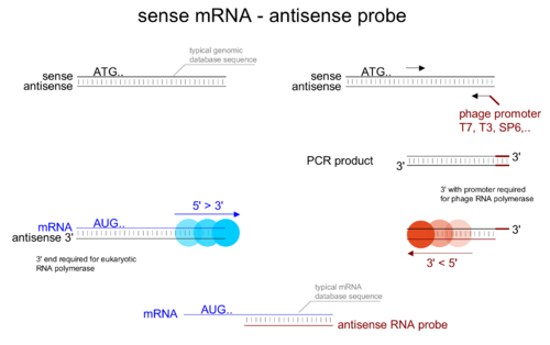
mRNA sense & antisense probes

Probes need to be antisense respective to the mRNA to be able to bind their target. Care has to be taken during experimental design to make a suitable antisense probe.


In vitro T7 transcription of an RNA probe

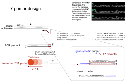
Designing primers for T7 RNA synthesis


  1. designing a suitable primer combining the T7 promoter sequence and a sequence specific part
  2. attaching a T7 promoter via PCR
  3. in vitro transcription using T7 polymerase and the PCR product as a template

Northern Blot, 32P End-Labeled Probes-PDF

Adapted from: Parker Lab Protocols, Sauer Lab, Heather Keller, Molecular Cloning, Current Protocols in Molecular Biology, Schleicher & Schuell Bioscience Turboblotter, Millipore Immobilon-Ny+ Transfer Membrane.

Contact: Caitlin Conboy

Contents

Samples to be probed

Experimental Samples

Total cellular RNA RNA Extraction ~10 µg of total cellular RNA, would contain 10 fg -1 pg of rare RNA, ~300 pg of moderately abundant RNA. (Molecular Cloning)

In Vitro Standard Curve Samples

RNA In Vitro Standards 1 fg – 100 pg range suggested. (Molecular Cloning) 3 – 50 ng for high copy/plasmid-based (cmc, 04-20-05)

Workflow outline

Probe Preparation

  1. For DNA probes: Design and order synthetic oligos (https://catalog.invitrogen.com/)
  2. End-Label Probe Prep, 32P End-Labeled Probes

Northern

  1. Gel
  2. Transfer
  3. UV Cross-linking
  4. Overnight Probe Hybridization
  5. Stringency Washes
  6. Incubation
  7. Phosphoimaging
  8. Strip/re-probe (optional)

**Radiation Use and Safety Reminders**

  1. Always record amount of radiation used per experiment in Radiation Log Book.
  2. Scan work area with Geiger counter before and after work.
  3. Always wear Radiation Badge when handling radioactivity.
  4. Use shields and radiation warning signs as necessary.
  5. Record activity of waste on waste container log sheets.

Protocol

Process the Gel (3-4 hours)

For RNase free electrophoresis apparatus: Clean electrophoresis tanks and combs used for electrophoresis of RNA with detergent solution, rinse in H20, dry with ethanol, and then fill with a solution of 3% H2O2. After 10 minutes at room temperature, rinse the electrophoresis tanks and combs thoroughly with H2O2 treated with 0.1% DEPC

  1. Set up the glyoxal denaturation reaction by combining 1-2 ul of RNA (up to 10 ug) with 10 ul of glyoxal reaction mixture. Run molecular weight markers for staining with sybrGold.
  2. Incubate the RNA solutions for 60 minutes at 55°C. Chill the samples for 10 minutes in ice water and then centrifuge them for 5 seconds to deposit all of the fluid in the bottom of the microfuge tubes.
  3. While the samples are incubating, clean electrophoresis tank if necessary, and pour a 1.5% agarose gel in 1X BPTE (1.05 g agarose in 70 mL buffer). When set, cover the gel with sufficient buffer.
  4. Add 2.5 ul of 6X gel loading buffer to the glyoxylated RNA samples, and without delay, load the glyoxylated RNA samples into the wells of the gel.
  5. Carry out electrophoresis at 70 Volts.
  6. Trim away areas of the gel to be stained with sybrGold. (Membrane should be cut to match the size of the gel.) Wrap gel to be stained in saran wrap and store at 4°C until post-transfer gel is ready to be stained.
  7. Soak gel 30 min in 0.05 M NaOH/1.5 M NaCl (~400 mL) without agitation.
  8. Soak gel 20 min in 0.5 M Tris (pH 7.4)/ 1.5 M NaCl (~400 mL)

7 and 8 are optional steps for improving the transfer of long RNAs, esp from >1% gels.

Prepare the Membrane (5-15 minutes)

NOTE: Be gentle with the membrane. The number of times a membrane can be stripped and re-probed is usually limited by physical damage to the blot.

  1. Cut a piece of membrane to the dimensions of the agarose gel. Max dimensions for hybridization in 50mL tubes: 8 x 9 cm (circumference x diameter). If membrane is larger, sandwich between sheets of nylon mesh to allow buffer to penetrate overlap.
  2. Wet the membrane by carefully laying it on top of Milli-Q water in a shallow tray. (Do not immerse the Immobilon-Ny+ membrane in liquid on the first liquid exposure. If you wet both sides, air can become trapped in the pores and form bubbles.)
  3. Agitate the tray gently once the membrane is wet to completely immerse the membrane.
  4. Transfer the membrane to a second tray containing transfer buffer (20 x SSC).
  5. Equilibrate the membrane at least 5 minutes.

Transfer of RNA onto Membrane by Turboblotter Capillary Transfer (3-4 hours)

NOTE: Refer to Fig. 1 when setting up the TurboBlotter System.

  1. Place stack tray of transfer device on bench, making sure it is level.
  2. Place 20 sheets of dry GB004 blotting paper (thick) in stack tray.
  3. Place 4 sheets of dry GB002 blotting paper (thin) on top of stack.
  4. Place one sheet of GB002 blotting paper, prewet in transfer buffer on stack.
  5. Place transfer membrane on stack.Smooth bubbles by rolling a clean glass pipette over the surface. Do not touch with gloves.
  6. Cover the membrane with agarose gel, cut the gel to the size of the membrane, making sure there are no air bubbles between the gel and the membrane.
  7. Wet the top surface of the gel with transfer buffer and place 3 sheets of GB002 Blotting Paper, presoaked in transfer buffer on top of the gel.
  8. Attach the buffer tray of the transfer device to the bottom tray using the circular alignment buttons to align both trays.
  9. Fill the buffer tray with 125 ml transfer buffer for 7 x 8 cm to 11 x 14 cm transfers; (200 ml for 12 x 21 cm to 20 x 25 cm transfers).
  10. Start the transfer by connecting the gel stack with the buffer tray using the precut buffer wick (included in each blotter stack), presoaked in transfer buffer. Place the wick cover on top of the stack to prevent evaporation. Make sure the edges of the wick are immersed in the transfer buffer.
  11. Continue the transfer for 3 hr. Additional transfer time may be required for gels thicker than 4 mm or larger-size nucleic acids. (Try 4 hr, since gel is 1.5%, cmc 3.25.05)
  12. Disassembly: mark edges of gel and lane borders onto blot with pencil.

NOTE: Do not place any other weight on top of the wick cover during transfer. This is unnecessary and may inhibit transfer by crushing the pore structure of the agarose gel.

RNA Fixation with UV Cross-Linking (5 minutes)

  1. It is not necessary to allow the blot to dry completely prior to UV cross-linking.
  2. Place the blot on a sheet of clean filter paper to prevent contamination if you plan to place the UV light source above the blotted RNA. (If you plan to place the membrane on a UV transilluminator, clean the surface with Milli-Q water and a Kimwipe.)
  3. Expose the side of the blot with the bound RNA to a UV light source (254 nm). Using Pabo Lab UV lamp, above blot, ~5 min. This lamp has not been calibrated, but multiple users have had success with 2-5 minutes exposure.

Hybridization (2 hours + overnight + >1 hour)

  1. If blot dimensions are less than 8 x 9 cm, place blot in a 50 mL conical tube, RNA facing in. Make sure the blot doesn’t overlap itself. 50 mL conical tubes fit in the Sauer Lab “Bambino” mini-hybridization oven. If blot is too large for a 50 mL conical tube, use large glass tubes and Baker Lab hybridization oven.
  2. Prewash Blot in 0.1x SSC/ 0.1% SDS for 1 hour at 65°C in hybridization oven. 10 – 15 mL of solution is required to cover the blot in a 50 mL tube, and sufficient for all steps. This incubation can be cut to 30 min if pressed (says Sean.)
  3. Remove Prewash
  4. Prehybridize blot for > 1 hour in 10 – 15 mL of pre-hybridization solution at hybridization temp. Hybridization temp ~15°C below estimated Tm of probe. If reusing probe in hybridization solution, thaw probe as balance. NOTE: It is possible to store blots in prehybridization solution sort-term to indefinitely at 4°C or -20°C (Sean)
  5. Remove prehybridization buffer if reusing probe, otherwise retain same buffer.
  6. Add hybridization buffer with old probe to blot or add new probe to pre-hybridization buffer. Hybridize >6 hrs at hybridization temp.
  7. Pour off probe either into 32P liquid waste (remember to record waste) or into tube for storage at -20°C for later reuse. NOTE: if storing probe in 13 mL conical tube, remember not to fill tube completely.
  8. Wash blot in 10 – 15 mL of 6x SSC/ 0.1% SDS for 5 min at room temp in the hybridization oven (Leave door open to change temp quickly. Put shield up.)
  9. Repeat wash twice for a total of three room temp washes. Dispose of the first wash in the 32P liquid waste. For subsequent washes pour off buffer in the sink. (Record waste.)
  10. During washes, pre-erase PhosphorImager screen 20 min on light table.
  11. Repeat wash a forth time for 20 min at 10°C below estimated Tm in hybridization oven.
  12. Pour off final wash. Remove damp blot from tube and lay on clean saran wrap. Fold saran wrap to seal blot.
  13. Expose wrapped blot to PhosphorImager screen in casette.
  14. Image screen after 1 hour.
  15. Erase screen.
  16. Expose screen overnight if original image is faint.
  17. Erase screen before returning casette.

Northern Blot Solutions

Prewash Solution (0.1x SSC/ 0.1% SDS)

50 mL Total Volume

    • 250 µL of 20x SSC
    • 500 µL of 10% SDS
    • 49.25 mL of nuclease-free H2O

100x Denhardt Solution

    • 5 g of Ficoll 400
    • 5 g of polyvinylpyrrolidone (Not polyvinylpolypyrrolidone!)
    • 5 g of BSA Fraction V (stored at 4°C)
  1. dissolve to 250 mL in nuclease-free H2O
  2. Sterile filter and freeze in 5 mL aliquots

Pre-hybridization Buffer

50 mL Total Volume

    • 5 mL of 100x Denhart’s Solution
    • 15 mL of 20x SSC
    • 0.5 mL of 10% SDS
    • 29.5 mL of Nuclease-free H2O

Stringency Wash (6x SSC/ 0.1% SDS)

200 mL Total Volume

    • 2 mL of 10% SDS
    • 60 mL of 20X SSC
    • 138 mL of nuclease-free H2O