Thursday, December 26, 2024
Home Blog Page 20

Preparing chemically competent cells-S2-PDF

Materials

  • Plate of cells to be made competent
  • TSS buffer
  • LB media
  • Ice

Glassware & Equipment

  • Falcon tubes
  • 500μl Eppendorf tubes, on ice
  • 200ml conical flask
  • 200μl pipeman or repeating pipettor
  • 5ml pipette

Preparation

  1. Grow a 5 mL seed culture of cells in LB medium to saturation. Dilute this culture back into 25–50 mL fresh LB in a 200 mL conical flask. You should aim to dilute the seed culture by at least 1/100.
  2. Grow the diluted culture at 30-37C and 250-400 RPM to an OD600 = 0.2–0.5. (You will get a very small pellet if you grow 25 mL to OD600 = 0.2)
  3. Put Eppendorf tubes on ice now so that they are cold when cells are aliquotted into them later. If your culture is X ml, you will need X tubes. At this point, you should also make sure that your TSS is being chilled (it should be stored at 4°C but if you have just made it fresh then put it in an ice bath).
    • 250 µL PCR tubes also work when aliquotting ≈50 µL cells. They save space and can be heated/cold-shocked on a thermocycler. 3 volume recovery medium can be added directly to the tube and incubated stationary without apparent efficiency loss (Dueber & Bennett Labs).
  4. Split the culture into two 50 mL falcon tubes and incubate on ice for 10 min.

All subsequent steps should be carried out at 4°C and the cells should be kept on ice wherever possible

  1. Centrifuge for 10 min at 3000 rpm and 4°C.
  2. Remove supernatant. The cell pellets should be sufficiently solid that you can just pour off the supernatant if you are careful. Pipette out any remaining media.
  3. Resuspend cells in chilled TSS buffer. The volume of TSS to use is 10% of the culture volume that you spun down. You may need to vortex gently to fully resuspend the culture, and keep an eye out for small cell aggregates even after the pellet is completely off the wall.
    • Higher concentrations of cells (2–3×) are found to enhance efficiency, requiring cell resuspension in 3–5% culture volume of TSS instead of 10% (Dueber & Bennett Lab).
  4. Add 100 µL aliquots to your chilled Eppendorf and store at -80°C.
    • The original paper [1] suggests freezing the cells immediately using a dry ice bath. I (BC) have used liquid nitrogen quite successfully instead of dry ice. Simply placing the cells at -80°C also seems to work well (Jkm).
    • If you run a control every time you clone (i.e. a vector-only ligation), you can also freeze cells in 200 µL aliquots. Unused cells can be frozen back once and reused, albeit with some loss of competence.
  5. It is a good idea to run a positive control on the cells.
    • The Endy Lab is trying to use a standard positive control to better compare (and hopefully improve) the transformation efficiencies in the lab, you can check it out here.

Notes

  • Note1: CT Chung’s paper recommends long storage of TSS competent cells at -70ËšC, while people have been using a wide range of temperatures from -20ËšC to -140ËšC for long-term storage.


Beta-Galactosidase Assay (A better Miller)-PDF

Background

β-Galactosidase is encoded by the lacZ gene of the lac operon in E. coli. It is a large (120 kDa, 1024 amino acids) protein that forms a tetramer. The enzyme’s function in the cell is to cleave lactose to glucose and galactose so that they can be used as carbon/energy sources. The synthetic compound o-nitrophenyl-β-D-galactoside (ONPG) is also recognized as a substrate and cleaved to yield galactose and o-nitrophenol which has a yellow color. When ONPG is in excess over the enzyme in a reaction, the production of o-nitrophenol per unit time is proportional to the concentration of β-Galactosidase; thus, the production of yellow color can be used to determine enzyme concentration.

So, why do we care? Usually, experiments are designed so that the β-Galactosidase concentration in the cell is a readout for some aspect of a system being studied. For example, an investigator may fuse a promoter to the lacZ gene and use β-Gal levels as a readout for promoter activity under various conditions. In 1972, Jeffrey Miller published “Experiments in Molecular Genetics” which contained a protocol for determining the amount of β-Gal with ONPG. Because of this, ONPG/β-Gal assays are referred to as “Miller” assays, and a standardized amount of β-Gal activity is a “Miller Unit”.

1 Miller Unit = [math]\displaystyle{ 1000 * \frac{(Abs_{420} – (1.75*Abs_{550}))}{(t * v * Abs_{600})} }[/math]

where:

  • Abs420 is the absorbance of the yellow o-nitrophenol,
  • Abs550 is the scatter from cell debris, which, when multiplied by 1.75 approximates the scatter observed at 420nm,
  • t = reaction time in minutes,
  • v = volume of culture assayed in milliliters,
  • Abs600† reflects cell density.

†Note that this value is different for each spectrophotometer used and should be calibrated by plating dilutions of known Abs600 cultures to determine the colony-forming units per Abs600.

In his book, Dr. Miller explains that this formula yields approximately 1 Miller Unit for uninduced E. coli (low β-Gal production) and approximately 1000 units for a fully induced culture (grown on lactose or IPTG).

In my experience, cultures of MG1655 induced with 1 mM IPTG in log phase have 1500-1800 Miller units. The reason for the difference is not known, but I suspect it stems from differences in the Abs600/cell density between Dr. Miller’s spectrophotometer and the one I use and the fact I do my Miller assays at 30 °C (for convenience) whereas Dr. Miller performed his assays at 28 °C. I have made promoter fusions that generate ~40,000 Miller units; however, as will be discussed below, this is too high for the assay and so the protocol was changed to lower this value.

Protocol

The protocol I use is derived from a paper by Zhang and Bremer (JBC 270, 1995, Free full text!) in which the original Miller protocol was greatly simplified to allow more samples to be measured with less manipulation.

In short, the protocol consists of measuring the cell density of a culture of bacteria (Abs600), then removing an aliquot of the cells from the cuvette and mixing them with a “permeabilization” solution that contains detergent which disrupts the cell membranes (but leaves the β-Gal intact). This kills the cells and stops translation. After incubation, an ONPG “substrate” solution is added and the yellow color allowed to develop. A “stop” solution is then added and the absorbance of o-nitrophenol is measured.

  1. Grow cultures under whatever conditions you wish to test.
  2. During growth, pre-measure 80 μL aliquots of permeabilization solution into 1.5 mL microfuge tubes and close them.
  3. Measure Abs600 and RECORD IT!
  4. Remove a 20 μL aliquot of the culture and add it to the 80 μL of permeabilization solution.

The sample is now stable for several hours. This allows you to perform time-course experiments.

  1. After the last sample is taken, move the samples and the Substrate solution to the 30 °C warm room for 20-30 minutes.
  2. Add 600 μL of Substrate solution to each tube and NOTE THE TIME OF ADDITION.
  3. After sufficient color has developed, add 700 μL of Stop solution, mix well, and NOTE THE STOP TIME.
  4. After stopping the last sample (some may take longer than others, but generally they are done in 30-90 minutes), transfer the tubes to a microfuge and spin for 5-10 minutes at full speed.
  5. Carefully remove the tubes from the centrifuge and transfer solution from the TOP of the tubes to your cuvette(s). You are trying to avoid having particulate material in the cuvette so that scattering will not influence the reading.
  6. Record Abs420. This should be less than 1 and greater than 0.05. If it’s a bit outside of this range, don’t sweat it.

Calculate Miller Units as:

[math]\displaystyle{ 1000 * \frac{(Abs_{420})}{((Abs_{600} \text{ of culture sampled})*(\text{volume } [0.02 \text{ mL}])*(\text{reaction time}))} }[/math]

Comments on the assay

  • Reshma 11:28, 15 October 2007 (CDT): Miller recommends a culture with OD600 = 0.28 to 0.70. However, he claims that overnight cultures can also be used but that exponentially growing cells give more precise assays.
  • When is the reaction done? I never found a good answer for this in the literature. If I let a reaction go to completion, I measured an Abs420 of ~2-3. Of course, this is out of the reliable range of the spectrophotometer, but it gives an indication of how far the reaction can go. I got reproducible data when the yellow color was just detectable before adding the stop solution up to about the color of LB broth before stopping. Remember, you need the substrate to saturate the enzyme during the course of the reaction, so don’t let them go too far. I routinely make three separate measurements for each culture and average them.
    • Reshma 11:28, 15 October 2007 (CDT): Miller recommends that the OD420nm reading should ideally be 0.6-0.9.
  • Frequently, people use disposable plastic cuvettes to measure both the culture turbidity and the yellow o-nitropenol. It is my experience that the disposable cuvetees have HORRIBLE optics: yes, they are clear, but the light path is pathetically distorted (just look through one at a distant object). This is not a big problem if the same cuvette is used for the blank and the sample and it is oriented in the same way in the spectrophotometer. The problem arises when many different samples are measured in different cuvettes. The values can can vary GREATLY even for the same culture measured in different cuvettes. I recommend using either a high quality glass or quartz cuvette, or measuring the samples in a plate reader using flat-bottomed 96 well plates. I have found my error in pipetting 150 μL of culture for Abs600 measurements was WAY less than using disposable cuvettes. Of course, the turbidity measured should be calibrated to cells/mL as with a 1cm cuvette.
  • By spinning the samples and carefully removing the a sample from the supernatant, you avoid having to measure the scattering at 550nm and guessing what it would be at 420nm.
  • If your reaction has too much β-Gal, the tube will turn yellow in a few minutes (or even seconds). This is too fast. One of the greatest contributions to error will be your estimate of reaction time. By having the reactions conditions set so that it takes about an hour, the time errors become insignificant. If you need to slow the reaction, you can use fewer cells and increase the amount of permeabilization buffer so the volume is still 100 μL. Alternately, you can re-engineer the ribosome-binding site of your β-Gal construct to weaken it. I found that if my cells were making 40,000 Miller units of β-Gal, they were very sick from the translation stress. It was better in this case to weaken the translation of the β-Gal mRNA.
    • I do these reactions like I do enzyme kinetics. I start the samples 10 sec apart (with the time counting up) so it is possible to get accurate reaction times even if you only let your reactions go for a few minutes. I get very reproducible results with reaction times of 1.5-30 min. Once you get a feel for how long your reactions will need, it is easy to group samples that will need the same reaction time. Doing each sample in triplicate will give you some confidence that your timing is reproducible.–Kathleen 16:43, 14 December 2005 (EST)
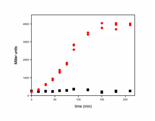
  • Here’s an example of some actual data I obtained using this assay. It was a timecourse experiment. At each time, I removed 1 mL from each of my cultures, measured the OD600, took three 20 µL aliquots directly from the cuvette and added each to 80 µL of permeabilization solution. I performed the assay exactly as described above, and all of the samples were kept at room temperature until the timecourse was finished. The OD600 for these cultures varied from 0.4 to 4 (in my spec) over the course of this experiment and the reaction times for the β-Gal assays varied from 2–25 min. The three individual β-Gal assays for each time point for each culture (red or black symbols) are plotted in the graph to illustrate the reproducibility of the assay within each experiment.–Kathleen

Recipes

Permeabilization Solution

You need 80 μL per sample.

  • 100 mM dibasic sodium phosphate (Na2HPO4)
    • (The Zhang protocol has 200 mM sodium phosphate. I could never get this into solution with the other components, no matter what I tried so I backed it off to 100 mM. I have even used 50 mM with no detectable change.)
  • 20 mM KCl
  • 2 mM MgSO4
  • 0.8 mg/mL CTAB (hexadecyltrimethylammonium bromide)
  • 0.4 mg/mL sodium deoxycholate
  • 5.4 μL/mL beta-mercaptoethanol

Substrate solution

You need 600 μL per sample.

  • 60 mM Na2HPO4
  • 40 mM NaH2PO4
  • 1 mg/mL o-nitrophenyl-β-D-Galactoside (ONPG)
  • 2.7 μL/mL β-mercaptoethanol

(The Zhang protocol also has 20 μg/mL CTAB and 10 μg/mL deoxycholate. I leave these out figuring that there is still plenty from the permeabilization solution and, if they ain’t dead yet, they ain’t gonna be.)

Stop solution

You need 700 μL per sample.

  • 1 M Sodium Carbonate (Na2CO3)

The high pH of the stop solution denatures the β-Gal and approximately doubles the yellow color of the reaction.


TOP10 chemically competent cells-PDF

0

Overview

This protocol is a variant of the Hanahan protocol [1] using CCMB80 buffer for DH10B, TOP10, and MachI strains. It builds on Example 2 of the Bloom05 patent as well. This protocol has been tested on TOP10, MachI, and BL21(DE3) cells. See Bacterial Transformation for a more general discussion of other techniques. The Jesse ‘464 patent describes using this buffer for DH5α cells. The Bloom04 patent describes the use of essentially the same protocol for the Invitrogen Mach 1 cells.

This is the chemical transformation protocol used by Tom Knight and the Registry of Standard Biological Parts.

Materials

  • Detergent-free, sterile glassware, and plasticware (see procedure)
  • Table-top OD600nm spectrophotometer
  • SOB

CCMB80 buffer

  • 10 mM KOAc pH 7.0 (10 ml of a 1M stock/L)
  • 80 mM CaCl2.2H2O (11.8 g/L)
  • 20 mM MnCl2.4H2O (4.0 g/L)
  • 10 mM MgCl2.6H2O (2.0 g/L)
  • 10% glycerol (100 ml/L)
  • adjust pH DOWN to 6.4 with 0.1N HCl if necessary
    • adjusting pH up will precipitate manganese dioxide from Mn-containing solutions.
  • sterile filter and store at 4°C
  • A slight dark precipitate appears not to affect its function
  • Note: you can buy pre-made CCMB80 buffer from Teknova

Procedure

Preparing glassware and media

Eliminating detergent

Detergent is a major inhibitor of competent cell growth and transformation. Glass and plastic must be detergent-free for these protocols. The easiest way to do this is to avoid washing glassware, and simply rinse it out. Autoclaving glassware filled 3/4 with DI water is an effective way to remove most detergent residue. Media and buffers should be prepared in detergent-free glassware and cultures grown up in detergent-free glassware.

Prechill plasticware and glassware

Prechill 250mL centrifuge tubes and screw cap tubes before use.

Preparing seed stocks

  • Streak TOP10 cells on an SOB plate and grow for single colonies at 23°C
    • room temperature works well
  • Pick single colonies into 2 ml of SOB medium and shake overnight at 23°C
    • room temperature works well
  • Add glycerol to 15%
  • Aliquot 1 ml samples to Nunc cryotubes
  • Place tubes into a zip lock bag, immerse the bag into a dry ice/ethanol bath for 5 minutes
    • This step may not be necessary
  • Place in -80°C freezer indefinitely.

Preparing competent cells

  • Inoculate 250 ml of SOB medium with 1 ml vial of seed stock and grow at 20°C to an OD600nm of 0.3
    • This takes approximately 16 hours.
    • Controlling the temperature makes this a more reproducible process, but is not essential.
    • Room temperature will work. You can adjust this temperature somewhat to fit your schedule
    • Aim for lower, not higher OD if you can’t hit this mark
  • Centrifuge at 3000rpm at 4°C for 10 minutes in a flat bottom centrifuge bottle.
    • Flat bottom centrifuge tubes make the fragile cells much easier to resuspend
    • It is often easier to resuspend pellets by mixing before adding large amounts of buffer
  • Discard supernatant by pouring out slowly and pipetting the remaining supernatant
  • Gently resuspend in 80 ml of ice-cold CCMB80 buffer
    • sometimes this is less than completely gentle. It still works.
  • Incubate on ice for 20 minutes
  • Centrifuge again at 4°C and discard supernatant as described above.
  • Resuspend in 10 ml of ice-cold CCMB80 buffer.
  • Test OD of a mixture of 200 μl SOC and 50 μl of the resuspended cells.
  • Add chilled CCMB80 to yield a final OD of 1.0-1.5 in this test.
  • Aliquot to chilled screw top 2 ml vials or 50 μl into chilled microtiter plates
  • Store at -80°C indefinitely.
    • Flash freezing does not appear to be necessary
  • Test competence (see below)
  • Thawing and refreezing partially used cell aliquots dramatically reduces transformation efficiency by about 3x the first time, and about 6x total after several freeze/thaw cycles.

Measurement of competence

  • Transform 50 μl of cells with 1 μl of standard pUC19 plasmid (Invitrogen)
    • This is at 10 pg/μl or 10-5 μg/μl
    • This can be made by diluting 1 μl of NEB pUC19 plasmid (1 μg/μl, NEB part number N3401S) into 100 ml of TE
  • Hold on ice 0.5 hours
  • Heat shock 60 sec at 42C
  • Add 250 μl SOC
  • Incubate at 37 C for 1 hour in 2 ml centrifuge tubes rotated
    • Using 2 ml centrifuge tubes for transformation and regrowth works well because the small volumes flow well when rotated, increasing aeration.
    • For our plasmids (pSB1AC3, pSB1AT3) which are chloramphenicol and tetracycline-resistant, we find growing for 2 hours yields many more colonies
    • Ampicillin and kanamycin appear to do fine with 1 hour growth
  • Plate 20 μl on AMP plates using sterile 3.5 mm glass beads
    • Good cells should yield around 100 – 400 colonies
    • Transformation efficiency is (dilution factor=15) x colony count x 105/µgDNA
    • We expect that the transformation efficiency should be between 5×10⁸ and 5×10⁹ cfu/µgDNA

5x Ligation Adjustment Buffer

  • Intended to be mixed with ligation reactions to adjust buffer composition to be near the CCMB80 buffer
  • KOAc 40 mM (40 ml/liter of 1 M KOAc solution, pH 7.0)
  • CaCl2 400 mM (200 ml/l of a 2 M solution)
  • MnCl2 100 mM (100 ml/l of a 1 M solution)
  • Glycerol 46.8% (468 ml/liter)
  • pH adjustment with 2.3% of a 10% acetic acid solution (12.8ml/liter)
    • Previous protocol indicated the amount of acetic acid added should be 23 ml/liter but that amount was found to be 2X too much per test on 1.23.07 –Meaganl 15:50, 25 January 2007 (EST)
  • water to 1 liter
  • autoclave or sterile filter
  • Test pH adjustment by mixing 4 parts ligation buffer + 1 part 5x ligation adjustment buffer and checking pH to be 6.3 – 6.5
  • Reshma 10:49, 11 February 2008 (CST): Use of the ligation adjustment buffer is optional.

References

  1. Hanahan D, Jessee J, and Bloom FR. Plasmid transformation of Escherichia coli and other bacteria. Methods Enzymol. 1991;204:63-113. DOI:10.1016/0076-6879(91)04006-a | PubMed ID:1943786 | HubMed [Hanahan91]
  2. Reusch RN, Hiske TW, and Sadoff HL. Poly-beta-hydroxybutyrate membrane structure and its relationship to genetic transformability in Escherichia coli. J Bacteriol. 1986 Nov;168(2):553-62. DOI:10.1128/jb.168.2.553-562.1986 | PubMed ID:3536850 | HubMed [Reusch86]
  3. Addison CJ, Chu SH, and Reusch RN. Polyhydroxybutyrate-enhanced transformation of log-phase Escherichia coli. Biotechniques. 2004 Sep;37(3):376-8, 380, 382. DOI:10.2144/04373ST01 | PubMed ID:15470891 | HubMed [Addison04]
  4. US Patent 6,709,852 Media:pat6709852.pdf[Bloom04]
  5. US Patent 6,855,494 Media:pat6855494.pdf[Bloom05]
  6. US Patent 6,960,464 Media:pat6960464.pdf[Jesse05]

All Medline abstracts: PubMed | HubMed

Miniprep/GET buffer-PDF

Reagents

  1. GET buffer = 50 mM glucose (MW 180), 10mM EDTA, 25 mM Tris-HCl pH 8
  2. Alkaline Buffer= 0.2 N NaOH
  3. Potassium acetate = 3 M potassium acetate, 1.8 M acetic acid, no pH adjustment
  4. SDS
  5. PCA solution (optional) = 50 parts phenol, 49 parts chloroform, and 1 part amyl-alcohol
  6. Lysozyme (optional)

Procedure

  1. Pipet 2 ml of overnight culture into a 2 ml centrifuge tube.
  2. Centrifuge for 1 minute at maximum speed and discard the supernatant.
  3. Add 100ul of refrigerated GET buffer to the pellet and vortex to resuspend.
  4. Freshly prepare the alkaline SDS solution by adding 10mg SDS per ml of 0.2M NaOH Solution.
  5. Add 200μl of alkaline SDS solution to the cell suspension and invert to mix. DO NOT VORTEX! The solution should become clear.
  6. Add 150ul of refrigerated potassium acetate solution and invert gently to mix. DO NOT VORTEX! A precipitate should form.
  7. Store the tube on ice for 3-5 minutes.
  8. Centrifuge for 10 minutes at maximum speed.
  9. Carefully pipet 400ul of the clean supernatant into a new tube. DO NOT PICK UP ANY PRECIPITATE!!!
  10. Add 900ul of 100% (95% is ok too) EtOH to precipitate the plasmid DNA.
  11. Place the tubes in the -80 freezer for 30 minutes.
  12. Centrifuge the precipitated plasmid DNA 15 minutes at maximum speed (cold if possible)and discard supernatant.
  13. Carefully add 1ml 70% EtOH to the pellet and let sit for 3 minutes.
  14. Centrifuge at maximum speed for 3 minutes. Make sure the pellet is toward the outside.
  15. Discard the supernatant and air dry the pellet for 10-15 minutes.
  16. Once the pellet is COMPLETELY DRY resuspend the plasmid DNA in 20 ul of TE or distilled water. The DNA will contain RNA contamination, which can be removed by resuspending in TE with RNAse.

Optional Steps

  • After step 3 add 10mg lysozyme and incubate for 30 mins before proceeding with step 5. This step is essential for lysing gram-positive cells.
  • After step 9 add 400µl PCA solution to the tube, invert to mix, and centrifuge for 3 minutes at maximum speed. Collect the upper phase by pipetting into a new tube and proceed with step 10. This step helps remove any residual proteins.

Notes

Scale-up

Even higher concentrations of plasmid have been obtained by scaling up this protocol 5x in 15ml centrifuge tubes. However, to do this a larger (i.e. slower) centrifuge was necessary to accomodate the larger tubes. The adjusted centrifugation steps were as follows:

Step 9: Centrifuge at 5500g (Max speed for this larger centrifuge) for 15 minutes.
Step 13: Centrifuge at 5500g for 20 minutes.


Maxiprep of plasmid DNA from E. coli-PDF

Ingredients

Ingredients are per culture; make enough for one extra culture to allow for pipetting error).

  • 150μL sterile 50% glycerol
  • 1mL TEG (25mM Tris-Cl, 10mM EDTA, 50mM dextrose)
  • 111μL 20mg/mL lysozyme
  • 2mL worth of components for solution 2: 200μL 10% SDS, 100μL 4M NaOH, 1.7 mL autoclaved water
  • 1.5mL Solution 3: 3M K+, 5M acetate (3M potassium-acetate, 2M acetic acid — glacial is 17M)
  • 3.7mL isopropanol
  • 1mL TE buffer
  • 0.5mL 5M LiCl
  • 7.5μL 1mg/mL RNaseA
  • 200μL 70% ethanol
  • several mL of phenol:chloroform:isoamyl alcohol (25:24:1)
  • several mL of chloroform:isoamyl alcohol (24:1)
  • 750μL straight ethanol
  • 125μL 3M sodium acetate

Directions

  1. Grow a single colony of E. coli overnight in 50mL LB broth + selective markers at 37°C.
  2. The next morning, put 850μL of the culture in each of two Eppendorf tubes, add 150μL sterile 50% glycerol, and store at -80°C. Pour as much culture as will fit into an Oak Ridge tube and centrifuge at 5800g/6000rpm, 4°C, for 10 minutes in a GSA rotor. Discard the supernatant, add the rest of the culture, and repeat. Resuspend in 1mL TEG.
  3. Add 111μL 20mg/mL lysozyme. Incubate on ice for 30 minutes. Meanwhile, mix: 250μL 10% SDS, 125μL 4M NaOH, 2.125mL autoclaved water per culture.
  4. Add 2mL SDS/NaOH mix to each tube. Incubate on ice for 10 minutes.
  5. Add 1.5mL Solution 3 (3M K+, 5M acetate). Incubate on ice for 10 minutes.
  6. Shake vigorously. Centrifuge in SS34 rotor at 17,200g/12,000rpm, 4°C, for 15 minutes.
  7. Pour the supernatant into another Oak Ridge tube and discard the pellet. Add 2.7mL isopropanol. Centrifuge at 17,200g/12,000rpm (room temperature) for 10 minutes. Discard the supernatant.
  8. Wash pellet with 1mL 70% ethanol. Air dry for 2-5 minutes on bench. Resuspend in 500μL TE buffer. Add 500μL 5M LiCl. Incubate on ice for 5 minutes.
  9. Centrifuge at 17,200g/12,000rpm for 10 minutes.
  10. Pour supernatant into an Eppendorf. Add 1mL isopropanol. Incubate on the bench for 10 minutes.
  11. Centrifuge at 17,200g/12,000rpm for 10 minutes.
  12. Discard the supernatant. Wash the pellets with 100μL 70% ethanol. Resuspend in 375μL TE buffer. Add 7.5μL 1mg/mL RNaseA. Incubate at 37°C for 30 minutes.
  13. Add 700μL phenol:chloroform:isoamyl alcohol. Vortex until thoroughly mixed. Centrifuge at top speed of microfuge for 2 minutes. Pipette aqueous phase (the top one) into new Eppendorf. Repeat until the interface between the phases is clear after centrifugation. Then repeat the procedure twice with chloroform:isoamyl alcohol to remove any phenol.
  14. Add 750μL straight ethanol and 125μL 3M sodium acetate. Put at -80°C for 30 minutes or -20°C overnight.
  15. Centrifuge at 13,600g/12,000rpm, 4°C, for 15 minutes. Discard the supernatant. Wash pellet with ~100μL 70% ethanol. Resuspend in 100-200μL TE buffer.

ChIP-Chip E. coli-PDF

Abstract

ChIP-Chip stands for Chromatin Immunoprecipitation and chip in the sense of DNA microarray. It is a technique to determine the genome-wide binding sites of a DNA-binding protein. While the basic principle is the same for all species, there are some differences in handling cells. This protocol is developed and tested for E. coli. It should work the same way for other bacteria but that remains to be proven. Published protocols also exist for other bacterial species, including Bacillus subtilis, Caulobacter crescentus and Mycobacterium tuberculosis.

Because E. coli can be grown to high cell densities relative to eukaryotes, it is possible to generate sufficient DNA to label without using a PCR-based method. This method uses strand displacement primer extension with Klenow DNA polymerase and amplifies the DNA ~10-fold, while simultaneously incorporating dye-coupled nucleotide.

Materials

  • 2 ml Ultralink protein A/G beads (catalog number 53132, Pierce)
  • Specific antibody for example RNA polymerase β subunit from Neoclone, Madison

Reagents

Material for about 50 chromatin immunoprecipitations:

  • 75 ml Formaldehyde (37%)
  • 500 ml 2.5 M Glycine
  • 1.5 l TBS (400 ml 5xTBS)
  • 50 ml TE
  • 20 mg/ml Proteinase K in TBS (store up to 1 year at -20 C)
  • 1 ml 100mM PMSF or 0.5 ml 250 mM Pefabloc


Lysis Buffer

Final concentration For 50ml
10mM Tris (pH 8.0) 500μL 1M
20% sucrose 10g
50mM NaCl 500μL 5M
10mM EDTA 1mL 0.5M
10 mg/ml lysozyme 0.5g
  • Note: Prepare the 50ml without lysozyme and than add the corresponing ammount to 10 ml aliquotes and store them ad -20°C


IP-Buffer (Immunoprecipitation Buffer)

Final concentration For 500ml
50 mM HEPES-KOH pH 7.5 50ml 0.5M
150 mM NaCl 15ml 5M
1 mM EDTA 1ml 0.5M
1% Triton X 100 5ml
0.1 % Sodium deoxycholate 0.5g
0.1 % SDS 5ml 10%


  • IP-Buffer with 500 mM NaCl: add 2.8mL 5M NaCl to 40mL IP-buffer


Wash-Buffer

Final concentration For 50ml
10mM Tris pH 8.0 500µl 1M
250 mM LiCl 5 ml 2.5M
1 mM EDTA 100µl 0.5M
0.5% Nonidet-P40 (=Triton X114) 250µl
0.5% Sodium deoxycholate 2.5ml 10%


Elution-Buffer

Final concentration For 10ml
50 mM Tris (pH 7.5) 500μL 1M
10 mM EDTA 200μL 0.5M
1% SDS 1mL 10%
ddH2O 8.3mL

For labeling the DNA:

  • BioPrime kit Invitrogen (18094-011)[1]
  • Cy3-dCTP GE Healthcare (PA53021)[2]
  • Cy5-dCTP GE Healthcare (PA55021) [3]
  • Deoxynucleotides (2mM dATP, 2mM dGTP, 2mM dTTP, 0.5mM dCTP mix). Prepared as follows:
    • 4μl 100mM dATP GE Healthcare (27-2035-01)
    • 4μl 100mM dGTP
    • 4μl 100mM dTTP
    • 1μl 100mM dCTP
    • 187μl Water
    • Store at -20 °C
  • QIAquick PCR purification columns Qiagen (28104)[4]

Arrays

  • Oxfod Gene Technology (ogt) provides good arrays in 4*44k format compatibel with Agilent scanner and hybridisation equipment [5]
  • Hybridisation buffer materials:
  • 5M Sodium chloride Sigma (S6316)
  • 12x MES prepared as follows:
    • 3.52g MES free acid monohydrate Sigma (M2933)
    • 9.66g MES Sodium salt Sigma (M3058)
    • Water to 40ml
    • pH to 6.5 with 1M Hydrochloric acid
    • Add water to 50ml
    • Filter sterilise with a 0.45μm filter
    • Store at 4 °C
  • 10% Triton prepared as follows:
    • 1ml Triton X100 Sigma (T8787)
    • 9ml water
    • Store at room temperature
  • 100% Formamide Sigma (F9037-100ml)
  • 0.5M EDTA Sigma (E7889)
  • 20x SSPE Sigma (S2015)
  • 10% N-Lauroylsarcosine prepared as follows:
    • 1g N-Lauroylsarcosine Sodium salt Sigma (L9150-100g)
    • Water to 10ml
    • Store at room temperature
  • Polyethylene glycol (PEG) 200 Sigma (Fluka) (88440)

Equipment

  • DNA Microarray Hybridization Chamber – SureHyb from Agilent [6]
  • DNA Microarray Hybridization Oven from Agilent [7]
  • DNA Microarray Scanner from Agilent [8]

Procedure

This protocol has been broken up into 3 “days” but for E. coli it is possible to perform the entire ChIP experiment in a single day as well as the amplification/labeling step (which can be done overnight).

Formaldehyde cross link and sonication:

  • 50ml culture in LB or AB medium at 30 or 37 °C until OD600 0.2
  • Add 27μl formaldehyde (37%) per ml medium (substract what you took out for messuring OD) => final concentration of about 1%
  • Shake slowly (100 RPM) for 20 min at RT
  • Add 10 ml of 2.5 M glycine => final concentration of about 0.5 M
  • Keep shaking for 5 min
  • Harvest 50 ml of cells for each DNA-preparation (centrifuge 2500 g, 4°C, 10 min)
  • Wash twice in cold 10 ml TBS (20mM; see Material) pH7.5

You can freeze the cell pellet and proceed later

  • Resuspend in 300 μl IP-Buffer with 1mM Pefabloc

Sonicate so that DNA fragments are on average about 500 bps (see “critical steps” section below). With a Bioruptor from Diagenode use 48 cycles of 30 s sonication and 30 s cooling. Note that sonication parameters will vary according to the sonicator and probe used. It is possible to use a cup-horn sonicator but this requires much longer sonication and it is important to ensure that sonication is even for all samples being sonicated.

  • Prepare two 2ml tubes with 1.5ml IP-Buffer each.
  • Add 150 µl of sonicated extract to each tube with IP-Buffer
  • Centrifuge 12,000 g, 4 °C, 10 min

IP:

  • Transfer 800 μl aliquots of the supernatant in 4 tubes for immunoprecipitation
  • Add 20 μl of a 50% slurry of protein A sepharose or protein A/G beads (note that the beads should be selected to work with the antibody being used)
  • Add specific antibody (for example 1 μl of RNA polymerase β subunit; see Material above)
  • Incubate at 4 °C overnight on a slow rotator or at room temperature for 90 minutes. Note that most antibodies work fine at room temperature but some require overnight incubation at 4 °C. This has to be determined empirically, as does the amount of antibody required for the IP.
  • Collect sephareose beads by centrifugation for 1 min at 3500 rpm
  • Pipett off supernatant and save as control DNA
  • All following wash steps should be on a rotator at room temperature for 3 min with 2 min centrifugation as above:
  1. Wash twice with 700 μl I-Buffer
  2. Wash with 700 μl I-Buffer with 500 mM NaCl
  3. Wash with 700 μl Wash-Buffer
  4. Wash with 700 μl TE
  • Add 100 μl of elution buffer. Gently pipet up and down two or three times in order to dislodge beads from the filter. Incubate 10 min in a 65 °C water bath. A water bath is used instead of other heating apparatuses in order to improve heat transfer.
  • Centrifuge beads 1 min at 3500 rpm, room temperature. Transfer supernatant of all 4 tubes into 1 new 1.5 ml tube.
  • Centrifuge tube for 1 min at 3500 rpm, room temperature to pellet remaining agarose beads.
  • Transfer supernatant into new 1.5 ml tube.
  • Add 340 μl TE and 4µl RNase A (20mg/ml)
  • Incubate for 90 min at 42°C
  • add 20 μl Proteinase K to each tube and divide on PCR tubes fitting your PCR machine (for example 4×100μl). (Note: many protocols no longer have this step; I get equivalent data when I skip this)
  • To reverse cross-links, place tubes into PCR machine. Incubate 2 hr at 42 °C, followed by 6 hr at 65 °C. If not using Proteinase K, incubate overnight at 65 °C, or boil samples for 10 minutes.
  • Purify DNA by phenol extraction and ethanol precipitation or using a PCR purification kit (e.g. from Qiagen)
  • Elute or resuspend in 10-20 ul water
  • Measure DNA-content ideally at a NanoDrop (should be around 0.2 to 0.4 μg)
  • Use 1 and 10 ng DNA as template for quantitative PCR with primers that are specific for a known binding site of your DNA-binding protein and one negative control

Isolation of control DNA:

  • control DNA is isolated from the supernatant of the IP reaction (see above)
  • Incubate 500µl of IP supernatant at 95°C for 10 min
  • Add 2 µl RNase A and incubate in waterbath at 42°C for 90 min
  • Extract DNA with 1 x phenole and 2 x chlorophorm
  • Precipitate with 1 ml ethanol and 40 μL 3M Na-Acetat and 1.5µl Glyco-Blue over night at -20°C
  • spin down DNA at 4°C for 15 min at max. speed, wash in 70% ethanol and resuspend pellet in 30 μL dH2O

Labeling for hybridisation to microarray:

  • The chromatin immunoprecipitated DNA samples should be in 20μl volume at a concentration of approximately 20ng/μl (more is also fine).
  • Note that one allways needs one test sample that will be labeld with one CyDye and a control labeld with another. As control one could use chromosomal DNA or DNA immunoprecipitated from a mutant strain of the DNA binding protein of choice or from different growth conditions etc.
  • add 20μL 2.5x Random primer (BioPrime kit) to each 20μl DNA sample.
  • Mix by flicking the tubes and spinning for 15 seconds in a microfuge.
  • Denature in a heat block at 94 degrees centigrade for 3 minutes.
  • Microfuge for 15 seconds.
  • Add the following to the tubes.
Test Sample 1(μl) Control Sample 2(μl)
dNTP mix (2mM dATP, 2mM dGTP, 2mM dTTP, 0.5mM dCTP) 5 5
Cy3-dCTP (1mM) 3.75
Cy5-dCTP (1mM) 3.75
Klenow (BioPrime kit) 1 1
  • Mix by flicking the tube followed by a brief (<10 secs) spin a microfuge.
  • Incubate at 37 °C for 5 hours (the time of incubation determins the degree of amplification so you could vary it if you want or need to).
  • Use QIAquick PCR purification kit for cleaning up the labeld DNA. Elute DNA with two times 25μL of elution buffer from the column. The colour of the column after the wash step gives a first impression of the degree of labeling.
  • Measure the CyDye and DNA concentration of the samples at Nanodrop. DNA should be between 20 and 60ng when started with about 20ng immunoprecipitated DNA and CyDye between 2 and 6 (just to give an idea).

Hybridisation to microarray

  • The following protocol is thought for hybridisation of OGT arrays with Agilent SureHyb equipment. If you want to use other equipment adjustments to this protocol may be required.
  • Remove slide box from packaging and store slides until use in a dehumidified chamber. The slides should be stored in a light tight box.
  • When ready for use, remove slides from box. Wear clean powder free gloves at all times when handling the microarrays. Handling should be carried out in a low dust laboratory. Return unused slides to dehumidified chamber.
  • The arrays are printed on the same side of the slide to the label ‘Agilent’.
  • Hybridisations are carried out in a 100μl volume per array.
  • The volume of CyDye labeld DNA must be reduced in a SpeedVac and be adjusted to 25μL.
  • Prepare the hybridisation buffer by pipetting the following into a tube. CARE Formamide is toxic.
Component Volume for 125μL hybridisation (μL) Volume for one slide with four arrays
12xMES 10 50
5M Sodium chloride 24 120
Formamide 24 120
0.5M EDTA 5 25
10% Triton X100 12 60
  • The given volumes in the first column are for one hybridisation. If you have more than one you should do a master mix (second column) and use 75μL in the following step.
  • In a different tube join the differentialy labeled test and control DNA as follows:
Component Volume for 125μL hybridisation (μL)
Cy3 labeld DNA 25
Cy5 labeld DNA 25
  • denature at 94°C for 3 min
  • Spin down and add sample mix in tube with hybridisation buffer (see above).
  • Place an Agilent SureHyb GASKET slide into an Agilent CHAMBER base.
  • Pipette 100μl of hybridisation mix onto the GASKET slide.
  • Place an OGT array slide onto the GASKET slide with the array side down (with ‘Agilent’ label) and in contact with the hybridisation mix.
  • Place the CLAMP ASSEMBLY on the slide and tighten the thumbscrew.
  • Some bubbles should form. These bubbles should be moving. If they are not, tap the chamber on the bench.
  • Hybridise at 55°C for 2 nights and one day (about 36 hours) in a light tight container, ideally in a hybridisation oven with a rotisserie. Fit the slides vertically and rotate the chambers at a speed at 4 rpm (setting 10 for Agilent hybridisation oven).

Washing and scanning of microarray

  • Note: Gloves should be changed after each wash step to not transfer CyDye.
  • Prepare the Wash solutions as follows
    • Wash 1 (1 litre)
    • 20x SSPE 300ml
    • 10% N-Lauroylsarcosine 0.5ml
    • Water 700ml

Store at room temperature.

  • Wash 2 (1 litre)
    • 20x SSPE 3ml
    • PEG200 1.8ml
    • Water 995ml
    • Store at room temperature.
  • Place 50ml of Wash 1 in a 50ml sterile tube.
  • Place 50ml of Wash 2 in a separate 50ml sterile tube.
  • Wearing gloves remove the slide from the hybridisation chamber with the GASKET slide still attached. CARE the hybridisation buffer contains formamide.
  • Place in a bath of Wash 1 and gently prise the GASKET slide from the OGT microarray under the surface of the Wash 1 buffer (use fingernails at the corners of the arrays).
  • Without the microarray drying out place the microarray into the 50ml tube with the Wash 1 buffer.
  • Rotate the tube on a rotary mixer at room temperature for 5 minutes.
  • Using clean forceps and without the microarray drying out, place the microarray into the 50ml tube with the Wash 2 buffer.
  • Rotate the tube on a rotary mixer at room temperature for exactly 5 minutes.
  • Using clean forceps remove the microarray and blow dry with dry nitrogen.
  • Insert the slide into the scanner and scan according the manufacturer’s instruction booklet. For Agilent scanner insert the slide into the Agilent slide holder. The array slide should be placed into the slide holder with the array side facing up. The ‘Agilent’ label should also be facing up. The non-labeled edge should be placed into the slide holder first. The slide should be scanned with the green laser (~532nm) and the red laser (~633nm).

Feature extraction with Agilent feature extraction software

  • An XML file is supplied from OGT on the CDc to enable the data to be extracted using Agilent’s Feature extraction software. Please refer to the Agilent Feature extraction software documentation for full details.
  • Carry out Feature extraction as recommended by the software provider
  • A .txt file should be generated. When the .txt file is opened using Microsoft Excel, a spreadsheet should open that will contain one column with the genomic location of the probe on the array. There will be another column with the Green and Red signals (gProcessedSignal and rProcessedSignal).
  • One could than for example draw a graph of the genomic location on the X axis versus the ratio of the two dye signals (test/control).
  • For OGT array 010010, the genomic location of the probes on the file is based on the EMBL E.coli K12 MG1655 genomic sequence (ID U00096).
  • For OGT array 010011, the genomic location of the probes on the 0157 array is based on the EMBL E.coli 0157 genomic sequence (ID BA000007). The plasmid sequence used is AB011549.
  • There is also a ChIP browser availible for free download from OGT [9] that helps to view ChIP data together with genomic location.

Critical steps

It is important to confirm that sonication results in fragments of a suitable size, i.e. <1 kb on average. This can be done by decrosslinking a sample of crosslinked cell extract, purifying by phenol extraction and ethanol precipitation, RNase treating and running on a gel.

Acknowledgments

Acnkowledge any help you had in development, testing, writing this protocol.

References

  1. Waldminghaus T and Skarstad K. ChIP on Chip: surprising results are often artifacts. BMC Genomics. 2010 Jul 5;11:414. DOI:10.1186/1471-2164-11-414 | PubMed ID:20602746 | HubMed [Waldminghaus-2010]


Engineering BioBrick vectors from BioBrick parts/Colony PCR-PDF

Materials

  • PCR SuperMix High Fidelity
  • VF2 primer (5'-TGCCACCTGACGTCTAAGAA-3')
  • VR primer (5'-ATTACCGCCTTTGAGTGAGC-3')
  • Deionized, sterile H2O
  • strip of PCR tubes
  • 2-log DNA ladder
  • 0.8% E-Gel® from Invitrogen Corporation in Carlsbad, CA

Equipment

  • DNA Engine OPTICON\texttrademark from MJ Research, Inc. (now Bio-Rad Laboratories, Inc., Hercules, CA)
  • E-Gel® PowerBaseâ„¢ v.4 from Invitrogen Corporation in Carlsbad, CA
  • Alpha Innotech FluoChemâ„¢ 880 gel imaging system

Procedure

PCR mix

  • 9 μL PCR SuperMix High Fidelity
  • 6.25 pmoles VF2 primer
  • 6.25 pmoles VR primer
  • 1 μL colony suspension
    • dilute 1 colony in 100 μL water

PCR conditions

  1. 95°C for 15 minutes
  2. 94°C for 30 seconds
  3. 62°C for 30 seconds
  4. 68°C for 3.5 minutes
  5. Repeat 2-4 39 times.
  6. 68°C for 20 mins
  7. 4°C forever

Agarose gel electrophoresis

  1. Dilute the reactions four-fold with water.
  2. Perform an agarose gel electrophoresis of 20 μL of each diluted reaction using a 0.8% E-Gel®.
  3. Also electrophorese 1 μg of 2-log DNA ladder to verify the length of each PCR product.
  4. Image gel with 302 nm transilluminating ultraviolet light using an ethidium bromide camera filter and an exposure time of 614 milliseconds.


Cell counting/plating-PDF

Overview

Count the number of viable cells in a culture via dilution plating. The following assumes E. coli cells but it should apply to any type of cells.

Materials

  • LB agar plate
  • LB media for dilution
  • 8-tube strips (optional but easiest)

Procedure

  1. Fill each tube in the dilution with 90 μl of LB
  2. Add 10 μl of the sample to the first tube and mix
  3. From the first tube, remove 10 μl and mix into second tube
  4. Repeat for the number of dilutions you wish to do (8 should be more than enough)
  5. Take 10 μl from each dilution and spot it on to the agar plate
  6. Allow droplet to dry and incubate

The first dilutions will contain a thick lawn of cells and the last dilutions will contain no cells. There should be one drop which contains countable single colonies. From this, you can calculate the number of cells in the original sample. For example, if there 4 colonies on dilution 5, there are 4E4 cells/μl.

STN Chemostat Protocol-PDF

Day 0

Set up o/n culture in supM9 media in a small flask at 37 C.

Day 1

  • Take OD600 of o/n culture. Dilute back in the morning into 25 mL/chamber supM9 (~1:100)
  • When culture reaches ~OD 2.0 to add 20 mL to each chamber (usually late afternoon).
  • Run at 37 C. The bubbler should be set to the white dot for 6 chambers (med/high) and the effluent needle should be set to around the 24 mL mark to maintain a volume of 20 mL.
  • Add the media input (teal, 23-G1 needle) and turn on pump (currently 10 rpm for 20 mL/chamber). After about 1 hour, check that the culture volume is 20 mL and adjust effluent as necessary.
  • The cultures should wash out to a stable OD600 by the morning (day 2). OD600 should be monitored throughout the day (~every 3 hrs) and samples drawn for steady-state measurement either in the latter part of day 2 or on day 3.

Notes

  • Run with bleach solution, then H20 after use. Autoclave all components on fast (22 psi, 250 F, 30 min.).
  • This water bath should be filled with dI. Top up frequently with dI kept at 37 C.
  • Use 1_5_05 supplemented M9 minimal media with AMP (50 ug/mL) or Kan (20 ug/mL) as needed.
  • Measure the flow rate by the effluent. 10 rpm gives about 0.25 mL/min, which is about right, but flow rate should be checked during each run.
  • Mark 18 mL, 20 mL, and 22 mL on chemostat chambers to assist in monitoring volume.

Cell cycle analysis-PDF

Overview

This protocol details the method for determining the initiation age (ai) and C+D period in bacterial cells, as well as the calculation of the C and D periods, the generation of a theoretical DNA histogram, and the calculation of the average number of replication forks.

Determination of initiation age (ai) and C+D:

From flow cytometry analysis of cells treated with rifampicin and cephalexin (run-out histogram) the proportions of cells that had not initiated replication at the time of drug action (4-origin-cells, streaked) and cells that had initiated (8-origin-cells) can be estimated.The initiation age (ai) can be found from the theoretical age distribution described by this formula,

[math]\displaystyle{ F=2-2^{\frac{(\tau-a_i)}{\tau}} }[/math]

where F is the fraction of cells that had not initiated and Ï„ is the generation time, or from the estimated graph of the theoretical age distribution (streaked portion).

This gives:

[math]\displaystyle{ a_i=\tau-\frac{log(2-F)}{log2}*\tau }[/math]

which is the same as this (log2 is 1):

[math]\displaystyle{ a_i=\tau-log(2-F)*\tau }[/math]

If you have for example a generation time Ï„=84 minutes and the portion of cells with 4 origins is 66% the formula gives:

[math]\displaystyle{ a_i=84-log(2-0.66)*84=48.5 }[/math]

The C+D period is estimated from the initiation age (ai), the generation time (Ï„) and the number of generations spanned per cell cycle.

Example:

4-origin-cells: 23 %

Generation time (Ï„): 27 min

Initiation age (ai): 5 min


Determination of the C and D periods:

The C period is found from the oriC/terC ratio obtained by Southern blot or qPCR analysis (oriC/ter ratio determination) and the generation time (Ï„):

[math]\displaystyle{ \frac{oriC}{terC}=2^{\frac{C}{\tau}} }[/math]

which gives:

[math]\displaystyle{ C=log_2(\frac{oriC}{terC})*{\tau} }[/math]

The D period is found from the C+D and C period:

[math]\displaystyle{ D = (C+D) – C }[/math]

Example (continues):

C period calculated from the oriC/terC ratio: 49 min

D period = (C+D) – C

D period = 76 min – 49 min = 27 min


The theoretical exponential DNA histogram:

A theoretical exponential DNA histogram can be drawn to check whether the obtained values fit with the experimental data. From the C+D period the DNA content of the cells at different time points in the cell cycle can be calculated.

Example:


The individual values of C and D can be varied

to obtain a shape of the theoretical histogram

that gives the best fit to the experimental histogram.

Calculation of the average number of replication forks when D=Ï„:

In the example given above, 23% of the cells contain 4 replication forks (4-origin peak in run-out histogram) and 77% contain 12 replication forks (8-origin peak), hence the average number of replication forks in the cell population will be:

(4 x 0.23) + (12 x 0.77) = 10.2 forks

Calculation of the average number of replication forks when D≠τ:

Example:

4-origin-cells: 23%

8-origin-cells: 77%

Ï„ = 27 min

ai = 5 min

C = 51 min

D = 25 min

C+D = 76 min


  1. 12 forks → 8-origin peak in run-out histogram = 77% of the cells
  2. 6 and 4 forks → 4-origin peak in run-out histogram = 23% of the cells
  3. The fraction of cells containing 6 forks: F = 2 – 2((Ï„-at)/Ï„) = 2 – 2((27-2)/27) = 0.10
  4. The fraction of cells containing 4 forks: 0.23 – 0.10 = 0.13
  5. The average number of replication forks: (6 x 0.10) + (4 x 0.13) + (12 x 0.77) = 10.4 forks