Friday, April 11, 2025
Home Blog Page 8

inclusion body solubilization-PDF

Buffers

buffer I (for purification on His-Trap columns)

  • 50 mM HEPES (pH 7.4)
  • 0.5 M NaCl (high salt)
  • 5 mM DTT (reducing conditions)
  • 8 M Urea (chaotropic, unfold proteins)
  • 1% v/v Triton X-100 (detergent, help solubilization of hydrophobic peptides)
  • 20 mM Imidazole (for reducing unspecific binding on the column)

Procedure

starting point: pellet of your insoluble fraction (after 40 min high-speed cold-room centrifugation of the lysate)

  1. add 500 µl buffer I to the pellet
  2. resuspend by pipetting or 2 times 5-10 s vortexing
  3. boil and load 5 µl + loading buffer on a SDS PAGE gel
  4. continue with purification using the same buffer without Triton and with varying amounts of Imidazole

in vitro FRET FRB FKBP-PDF

Overview

This protocol describes how to measure FRET efficiencies for a chemically induced interaction between FRB and FKBP12. A first measurement is done without rapamycin. Then rapamycin is added in three-fold excess. Sensitized (acceptor) emission is measured with an excess of donor (0.3µM acceptor + 0.5µM donor). Donor quenching is measured with an excess of acceptor (0.5µM donor + 0.3µM acceptor).

Materials

  • 1x HBS-P+ with 50 µM EDTA — our standard measurement buffer
  • fluorescence plate reader
  • black 96-well flat-bottom plates
  • multi-dispensing pipette

Procedure

Sensitized emission

Final conditions: 0.5µM donor + 0.3µM acceptor + 1.5µM Rapamycin in HBSP+

plate layout (5 or 6 replicas each):

  • row A: 150µl blank
  • row B (D): 50µl buffer + 100µl donor
  • row C (A): 100µl buffer + 50µl acceptor
  • row D (A+D): 100µl donor + 50µl acceptor

prepare solutions: (volumes are for one set of 6 replicas)

  1. dilute donor protein to 0.75µM in HBSP+; V=1400µl (1200+reserve)
  2. dilute acceptor protein to 0.9µM in HBSP+; V=720µl (600+reserve)
  3. dilute Rapamycin to 112.5µM; V=100µl (50+reserve)
  4. adjust plate reader (set excitation to donor absorption, and emission to acceptor emission peak)

prepare plate:

  1. multi-dispense buffer:
    • row A (blank): 150µl buffer
    • row B (donor): 50µl buffer
    • row C (acceptor): 100µl buffer
  2. multi-dispense donor (D):
    • row B: 100µl
    • row D: 100µl
  3. multi-dispense acceptor (A):
    • row C: 50µl
    • row D: 50µl
  4. mix plate by shaking 10s @ 1200 r.p.m.

measure w/o, with Rapamycin:

  1. load plate and measure acceptor emission after donor excitation for all wells
  2. remove plate
  3. multi-dispense 30µl rapamycin into a PCR stripe
  4. copy 2µl rapamycin into each well using a multi-channel pipette
  5. mix plate by shaking 10s @ 1200 r.p.m.
  6. repeat measurement twice

Donor quenching

Final conditions: 0.3µM donor + 0.5µM acceptor + 1.5µM Rapamycin in HBSP+

plate layout — same as above (5 or 6 replicas each):

  • row A: 150µl blank
  • row B (D): 50µl buffer + 100µl donor
  • row C (A): 100µl buffer + 50µl acceptor
  • row D (A+D): 100µl donor + 50µl acceptor

prepare solutions:

  1. dilute donor protein to 0.45µM in HBSP+
  2. dilute acceptor protein to 1.5µM in HBSP+
  3. dilute Rapamycin to 112.5µM
  4. adjust plate reader (set excitation to donor absorption, and emission to donor emission peak)

Now follow the remaining steps of the sensitized acceptor measurements.

Analysis

to be described.

Notes

Please feel free to post comments, questions, or improvements to this protocol. Happy to have your input!

raik:share your experience!

Polar Lipid Analysis-PDF

Polar Lipid Analysis

From DSM Manual, courtesy Hans-Juergen Busse.

Extraction and analysis of respiratory lipoquinones.

A number of methods have been described for the extraction of respiratory lipoquinones from microorganisms. These include the use of acetone, mixtures of chlorform:methanol (2:1 v/v) or hexane:methanol (1:2 v/v). Extraction is usually carried out using freeze dried cell material, although freshly harvested or frozen cell material is used by some groups. In order to avoid loss of lipoquinone material it is avisable to carry out the extraction under nitrogen atmosphere, away from strong sunlight, and at temperatures below 35-40 C. The method described is routinely used int he author’s laboratory, and permits the subsequent extraction of polar lipids from the same biomass.

1. 100 mg of freeze dried cell material is weighed into small screw cap bottles (10 ml capacity, brown glass) fitted with teflon-coated septa.

2. A small stirring bar is made from a 1 cm long piece of paper clip an also placed in the bottle.

3. To the cell material in the bottle is now added 3 ml of hexane:methanol (1:2 v/v).

4. Briefly gas the cell suspension with nitrogen and seal the bottle with the screw cap, making sure that the teflon-coated side seals against the mouth of the bottle.

5. The bottle is placed on a magnetic stirrer (about 10 such bottles can be placed on the stirrer at once) and left to stir for 30 min.

6. After 30 min stirring under nitrogen, the extracted material is placed in an ice bath until the hexane and methanol phases begin to separate.

7. Ice cold hexane (1 ml) is then added to give a methanol:hexane (1:1 v/v) biphasic mixture. Separation of the layers is enhanced by centrifuging the suspension in glass centrifuge tubes at 3000 rpm for 5 min.

8. The upper, hexane, phase is removed with a pasteur pipet and placed in a small glass tube (4-5 ml capacity).

9. The methanol phase is further extracted by the addition of cold hexane (2 ml) and 0.3% NaCl (2 ml) to give a 1:1:1 (v/v/v) ratio of hexane, methanol, and 0.3% NaCl. Centrifuge as above.

10. The upper hexane layer is then removed and added to the first hexane fraction.

11. The hexane phase is concentrated under a stream of nitrogen to give a final volume of about 0.5 ml, and the extract applied as a thin line to the lower edge of a silica gel plate (containing a fluorescent indicator F254).

12. The plates are developed in hexane:tert-butylmethylether (9:1 v/v) until the solvent front reaches the upper edge of the plate (20-30 min).

13. The developed plate is examined under UV light (254 nm) briefly to locate respiratory lipoquinones, which appear as dark bands against a greenish background. Menaquinones have an Rf value of about 0.7, while ubiquinones have an Rf of about 0.4.

14. The lipoquinone material is removed from the silica gel by scraping the appropriate area from the thin layer plate and placing it in a narrow bore tube (or pasteur pipet) fitted with a frit or glass wool plug. After tapping the tube to settle the silica gel, the lipoquinone material is eluted by passing 2x 0.5 ml of hexane:methanol (1:2 v/v) through the column, the eluant being collected in a small glass bottle (1.5 ml capacity). The purified lipoquinones may be recovered into hexane by cooling the mixture until phase separation occurs, followed by the addition of a few drops of 0.3% NaCl and about 0.3 ml of cold hexane. The upper, hexane phase containing the lipoquinones, is transferred to a second small 1.5 ml glass bottle, and may be stored at -20 C for analysis by UV spectroscopy, HPLC, or mass spectroscopy.

15. Further analysis of the lipoquinones is carried out by evaporating the hexane phase. HPLC analysis of the lipoquinones is carried out by dissolving the material in 100-200 ul of the HPLC solvent. UV spectroscopy is carried out by dissolving eh lipoquinone in a suitable volume (usually 0.5 – 1 ml) of spectroscopic grade hexane, ethanol, or (more commonly) iso-octane. Mass spectra of lipoquinones are recorded on material dissolved in the minimum quantity of a suitable solvent, such as methanol.

UV Spectroscopy

UV Spectra of respiratory lipoquinones give information on the nature of the quinone nucleus. UV spectra may be recorded in hexane, ethanol or iso-octane (the latter being the most frequently used). Spectra are recorded between 200 nm and 500 nm in quartz cuvettes. Menaquinone type lipoquinones (naphthoquinones) give four maxima between 230 nm and 280 nm, while ubiquinone type lipoquinones (benzoquionones) give two maxima lying very close together between 260 nm and 280 nm.


Polar Lipid Extraction

Polar lipids are generally extracted from dry cell material using chloroform:methanol:0.3% NaCl (1:2:0.8 v/v/v). This may be carried out by adding 9.5 ml of this mixture to 100 mg of freeze dried cells, or by adding a suitable amount of chloroform, methanol and 0.3% NaCl to the cell material, or to the aqueous methanolic phase remaining from the lipoquinone extraction.

1. The aqueous methanolic phase (4 ml total volume), together with the cell material from the lipoquinone analysis, is diluted with 5.5 ml of Chloroform:Methanol (2.5:3.0 v/v) to give a chloroform, methanol, 0.3% NaCl (1:2:0.8 v/v/v) mixture.

2. The mixture is placed in a 15 ml bottle with a teflon lined screw cap (check that the magnetic stirrer is in the bottle), gassed briefly with nitrogen, sealed and heated for 15 min at 80 C (with occasional shaking).

3. Allow the mixture to cool to room temperature on a magnetic stirrer. Check that the mixture is homogeneous, the presence of excess hexane will cause phase separation and may be overcome by adding a small amount of methanol until a homogeneous mixture is obtained.

4. The cell debris is removed by centrifugation in glass centrifuge tubes, at 3000 rpm for 5-10 min, and the supernatant decanted into 5 ml of chloroform:0.3% NaCl (1:1 v/v). The latter mixture (2.5 ml chloroform and 2.5 ml of 0.3% NaCl) may be placed in the extraction buffer bottles after they have been washed with distilled water.

5. After brief mixing, the biphasic mixture is centrifuged in glass centrifuge tubes (3000 rpm for 5 min) and the lower, chloroform phase collected with a pasteur pipet. Take care not to remove the flocculent protein layer or the aqueous phase.

6. The chloroform phase, containing the polar lipids is taken to dryness under a stream of nitrogen (when using a heating block or a water bath to accelerate drying, do not exceed 40 C).

7. The dried lipid material is then re-dissolved in 250 ul of chloroform:methanol (2:1 v/v) and transferred toa small glass bottle or ampule and may be stored for periods of several months at temperatures of -20 C or lower.

8. The lipid solution is used to spot thin layer plates; the lipids being placed in the bottom left-hand corner of each plate (Fig 4-A). Glass or aluminum plates are used because many detection reactions require the plates to be heated.

Run the first dimension with the spotted lipid on the lower right hand corner. After drying, run the plates in the second dimension with the lipid in the lower left hand corner.

9. Develop the plates in two dimensions using, in the first dimension chloroform:methanol:water (65:25:4 v/v/v), and in the second dimension, chloroform:methanol:acetic acid:water (80:12:15:4 v/v/v/v). Between the two dimensions, the plates should be dried at room temperature for about 20-30 min.

10. Lipid functional groups are identified using spray reagents specific for phosphate (Zinzadze), alpha-glycols (periodate-Schiff), and sugars (alpha-naphthol/H2SO4, anisaldehyde/H2SO4), free amino groups (ninhydrin), quaternary nitrogen (Dragendorff), and primary and secondary amines (chlorine and starch/iodide). Identification of the various lipids is carried out on the basis of staining reaction and Rf values. All spraying must be carried out in a fume hood using sufficient ventilation, since all lipid spray reagents are toxic.

Dual Immunofluorescence Cell Staining-PDF

Dual Immunofluorescence Labeling Protocol

Dual labeling assay using NUNC Labtek II chamberslide system (Fisher Scientific). For this assay, system was HA epitope-tagged (N-terminal) human P2Y2 retrovirally (pLXSN) infected into adherent 1321N1 human astrocytes that were stably maintained in DMEM 5% FBS, penn/strep and 500 ul/ml G418 (selection). Entire assay performed in a sterile tissue culture hood with minimal light-fluorescent or natural.

Procedure

  • Grow cells on labtek II slides to subconfluency (70%) at which point wash them and replace with serum free media for overnight (for purinergic receptors only; add 2 units/ml of a-pyrase in ddH20 to the starvation media solution to minimize receptor activiation/internalization due to secreted nucleotide phosphates)

  • Prior to starting the first labeling incubation prepare 40 ml 0.5% triton X100 per 2 slides and 40 ml 1% formalin phosphate / 2 slides in beakers or trays. The actual anitbody incubations will take place in 100 x 100 mm square multiuse trays (blotting trays) so you will need to cut out square pieces of filter paper and wet them with ddH20 and place them in the 100 x 100 um trays to maintain a humid environment during the antibody exposure. Prepare 40 ml DMEM +G418 (or whatever media) per two slides and warm to 37 degrees celsius.

  • Work with two slides at a time. Prepare one serum free DMEM wash in a 100 ml beaker. The first anitbody incubation is high affinity anti-HA rat monoclonal (12CA5) used at 0.1ug/100 ul. It takes approximately 1 ml of antibody solution (DMEM and G418 no serum or antibiotics) to assay 2 slides. Prepare the antibody solution and subsequently remove the wells using the tray removal tool (dump out media first). Work swiftly to prevent cells from drying. Dip the entire slide gently into the media wash. Gently remove and shake to release residual media. Place the slide into the 100 x100 mm tray. (Preheated to 37 degress with dH20 moistened filter paper) and gently add antibody solution. I used 300 ul per 8 well labtec slide applying 150 ul at a time. Incubate in 37 degree culture chamber for sixty minutes.

  • Five to ten minutes prior to the end of the first anitbody incubation, prepare your secondary (fluorochrome) anitbody labeling setup. This includes three washes in 100 ml beakers, two PBS washes and one serum free media wash and a clean 100 x 100 mm multiuse tray preheated with new moistened filter paper. Take slides that have been incubated for sixty minutes with HA probe and do the sequential washes one slide at a time. Lift the slide and gently dip and very gently swirl in PBS washes 3-5x, then finaly the DMEM (culture media wash) 3-5x and add the next antibody (in same fashion as the first) which is the fluoresent conjugate directed against HA probe (in this case oregon green 488 (molecualr probes), (1:100). Place the slides in a small light proof box. Incubate this anitbody for sixty minutes at 37 degress in culture chamber.

  • Five to ten minutes prior to the end of the second antibody incubation, set up three washes in 100 ml beakers, two PBS washes and one serum free media wash. Wash the slides as in step 4, but now place your slides (you want them to be completely submerged) in DMEM serum free in 100 x100 mm trays, place in light proof box, and allow to equilibrate 60 minutes in culture chamber.

  • You are now at the assay step. basically the cells are living at this point and have a flourescent chromophore label on the receptor via the HA probe and have been reequilibrated. To assay P2Y2, I used agonist concentration 100 uM UTP in DMEM serum free. I will assay an entire slide, either negative or positive UTP to keep the assay as controlled as possilbe (while surface tension between well dividers is sufficient to prevent bleeding of drops on the glass there is the potential for bleeding which is not good if you try to assay and + and – on a single slide). After three minutes exposure to 100 uM UTP, arrest the cells by placing the slide in 4 degree ice cold, 1% formalin PBS for ten minutes. After fixation, vacuum out formalin solution and replace with ice cold 0.5% triton x-100 for one minute to permeabilize cells. Subsequently vacuum out triton and add TBS. Prepare 2 TBS washes and the next antibody solution (1:100 in 3% BSA (EGFR sc-03 SantaCruz)). Wash slides and add next anitbody. Place in 100 x 100 mm tray as preformed in steps 3 and 4. This time incubate in this box and place box in a dark area at r.t. for 60 minutes.

  • After incubation, wash the slide in three separate 100 ul baths of TBS and add the second flouresenct conjugate, in this case anit-rabbit CY5 conjugate (Jackson Immunoresearch), 1:100. Incubate r.t. for 60 minutes. Note: Immediately, after you begin this final incubation, prepare the antifade reagent for fixing the slides. ***Prolong antifade kit by Molecular Probes is a good kit to use.

  • 5-10 minutes prior to the final incubation prepare 3×100 ml TBS washes and one large 500 ml ddH20 wash. Gently wash the slides through TBS and final water rinse and prop at an angle to allow them to partially dry. (You will see liquid evaporate slowly off slide when faced up. Wait for all liquid to evaporate and add preparative.) Use 25 x 25 mm cover slips and apply approximately 5 ul drop (no air bubbles) to each area (8 areas for 8 well plates) Gently drop the coverslip onto the slide and align. Allow it to settle. Place fixed slide in light proof box and place in 4 degree overnight prior to performing the visualization.

  • Assay under confocal scope.

The mounting agent is two component, one powder and one liquid. When you add the liquid make sure all the powder is dissolved. The powder tends to be in a chunks on the bottom of the the jar vial. Vortexing may not loosen up this chunk and into its dissolved state. Also let the soultion set for 5 minutes before pipeting to allow air bubbles to rise out of the glycerol. Molecular Probes Prolong Antifade Kit

Resources

Reference

Puromycin Selection-PDF

Puromycin dihydrochloride

Puromycin dihydrochloride is a aminonuclease antibiotic that inhibits protein synthesis. Puromycin is used for selection and maintenance of cell lines expressing a transfected pac gene (S. alboniger), whose product, puromycin acetyltransferase, inactivates puromycin via acetylation; recommended concentration in cell culture 1-10µg/ml. Lot-to-lot variations in potency exist for all selection antibiotics, each new lot of puromycin should be titrated. The working puromycin concentration for mammalian cell lines ranges from 1-10 µg/ml. Prior to using the puromycin antibiotic, titrate the selection agent to determine the optimal concentration for target cell line. Use the lowest concentration that kills 100% of non-transfected cells in 3-5 days from the start of puromycin selection.

Depending on individual cell type and doubling rate, selection of stable transfectants will take between 7 and 28 days. Expansion and characterization of single cell clones will take several weeks in addition. Media should be changed out every 2-3 days. This eliminates potentially toxic substances produced by dying cells and secondly, it keeps the concentration of the antibiotic at a constant level.

Streptomyces alboniger Puromycin N-acetyltransferase P13249

puromycin N-acetyltransferase

ATGACCGAGTACAAGCCCACGGTGCGCCTCGCCACCCGCGACGACGTCCCCAGGGCCGTACGCACCCTCGCCGCCGCGTTCGCCGACTACCCCGCCACGCGCCACACC GTCGATCCGGACCGCCACATCGAGCGGGTCACCGAGCTGCAAGAACTCTTCCTCACGCGCGTCGGGCTCGACATCGGCAAGGTGTGGGTCGCGGACGACGGCGCCGCG GTGGCGGTCTGGACCACGCCGGAGAGCGTCGAAGCGGGGGCGGTGTTCGCCGAGATCGGCCCGCGCATGGCCGAGTTGAGCGGTTCCCGGCTGGCCGCGCAGCAACAG ATGGAAGGCCTCCTGGCGCCGCACCGGCCCAAGGAGCCCGCGTGGTTCCTGGCCACCGTCGGCGTCTCGCCCGACCACCAGGGCAAGGGTCTGGGCAGCGCCGTCGTG CTCCCCGGAGTGGAGGCGGCCGAGCGCGCCGGGGTGCCCGCCTTCCTGGAGACCTCCGCGCCCCGCAACCTCCCCTTCTACGAGCGGCTCGGCTTCACCGTCACCGCC GACGTCGAGGTGCCCGAAGGACCGCGCACCTGGTGCATGACCCGCAAGCCCGGTGCCTGA

Puromycin Selection post transfection (shRNA)/transduction (lenti)

  • 48 hours post-shRNA transfection, aspirate the medium and replace with fresh medium containing puromycin at the appropriate concentration.
  • Approximately every 2-3 days, aspirate and replace with freshly prepared selective media.
  • Monitor the cells daily. Puromycin selection requires a minimum of 48 hours.
  • Optimum effectiveness should be reached within 3-10 days.
  • Assay transfected cells.

Suggested working conditions for selection in some mammalian cells:

  • Hela human uterus: 3 µg/ml
  • HEK293 human embryonic kidney: 3
  • B16 mouse melanoma: 1-3
  • PC1.0 hamster adenocarcinoma: 10
  • HEK293: 2
  • A431: 1
  • Jurkat: 4

Titrating puromycin

  • Plate 2 x 10⁵ cells in each well of a 6-well plate containing 3 ml of the appropriate complete medium plus increasing concentrations of puromycin (i.e., 0, 1.0, 2.5, 5.0, 7.5, and 10.0 µg/ml)
  • Replace with fresh selective medium after 2 days to remove dead cells.
  • Examine the wells for viable cells every two days.
  • Monitor the cells daily and observe the percentage of surviving cells. Optimum effectiveness should be reached in 1-4 days.
  • The minimum antibiotic concentration to use is the lowest concentration that kills 100% of the cells in 3-5 days from the start of puromycin selection.

Factors Influencing Successful Transfection

  • Concentration and purity of nucleic acids – Determine the concentration of your DNA using 260 nm absorbance. Avoid cytotoxic effects by using pure preparations of nucleic acids.
  • Transfection in serum-free media – the highest transfection efficiencies can be obtained if the cells are exposed to the transfection complexes in serum free conditions followed by the addition of medium containing twice the amount of normal serum to the complex medium 3–5 hrs post transfection (leaving the complexes on the cells). However, the transfection medium can be replaced with normal growth medium if high toxicity is observed.
  • No antibiotics in transfection medium – The presence of antibiotics can adversely affect the transfection efficiency and lead to increased toxicity levels in some cell types. It is recommended that these additives be initially excluded until optimized conditions are achieved, then these components can be added, and the cells can be monitored for any changes in the transfection results.
  • High protein expression levels – Some proteins when expressed at high levels can by cytotoxic; this effect can also be cell line specific.
  • Cell history, density, and passage number –It is very important to use healthy cells that are regularly passaged and in growth phase. The highest transfection efficiencies are achieved if cells are plated the day before. However, adequate time should be allowed to allow the cells to recover from the passaging (generally >12 hours). Plate cells at a consistent density to minimize experimental variation. If transfection efficiencies are low or reduction occurs over time, thawing a new batch of cells or using cells with a lower passage number may improve the results.

Measuring Puromycin cytotoxic

The measurement of cell viability and growth is an important factor in titrating antibiotic. Several approaches exist;

  • Trypan blue; live cells or tissues with intact cell membranes are not colored. Since cells are selective in the compounds that pass through the membrane, in a viable cell trypan blue is not absorbed; however, it traverses the membrane in a dead cell.
  • Bright field microscope can indicate adherent cells exhibiting loss of plate attachment
  • MTS assay water-soluble formazan product with absorbance maximum at 490-500 nm in PBS takes place only when reductase enzymes are active, and therefore conversion is a function of viable (living) cells.

References

Amaxa

Open Biosystems

  1. Murnane JP, Yezzi MJ, and Young BR. Recombination events during integration of transfected DNA into normal human cells. Nucleic Acids Res. 1990 May 11;18(9):2733-8. DOI:10.1093/nar/18.9.2733 | PubMed ID:2339059 | HubMed [Paper1]
  2. Haber JE. DNA repair. Gatekeepers of recombination. Nature. 1999 Apr 22;398(6729):665, 667. DOI:10.1038/19423 | PubMed ID:10227286 | HubMed [Paper2]
  3. Glover DJ, Lipps HJ, and Jans DA. Towards safe, non-viral therapeutic gene expression in humans. Nat Rev Genet. 2005 Apr;6(4):299-310. DOI:10.1038/nrg1577 | PubMed ID:15761468 | HubMed [Paper3]

All Medline abstracts: PubMed | HubMed

shRNA Transfection-PDF

Lentivirus, shRNA plasmid, or siRNA?

Choosing between Lentivirus (somewhat stable), shRNA transfer vector (stable/transient), or siRNA (transient). Perform and exhaustive literature search (pubmed) for citations that have reported RNA/DNA delivery methodology to (species) (lineage) cells. Discuss, evaluate and compare the materials and methods.

RNA (siRNA)

  • For easy to transfect cell line, RNA/DNA delivery protocol/procedure should be well defined. siRNA is a proven and effective method targeting mRNA as a molar calculable substrate. In studying effects that can be measured with transient mRNA knockdown, siRNA is a reliable and straightforward approach. Empirical moles of duplex/# cells or total volume (molarity) combines with a minimal number of steps and peripheral controls toward achieving results.

DNA (shRNA)

  • shRNA transfer vectors are ~7 kb DNA constructs that will be more stable to handle than RNA. However there are considerable nuances in the actual design and cloning of these vectors, in the actual establishment of antibiotic resistance toward generating a stable population, and in controlling for specific silencing/reproducible results.
  • shRNA is a large delivery vector producing a small hairpin substrate over cell passages under antibiotic selective pressure (ie puro resistance may not = Pol III shRNA cassette expression).
  • shRNA transfer vector alone may work insofar as culturing the cell, liposomal DNA transfection, antibiotic selection, and then collect data. However the lentivirus transfer vector is engineered for packaging into transducible (VSV-G) pseudotyped viral particles.

Lentivirus

  • For primary cell and/or differentiation restrictive (neuron), transducible (VSV-G) pseudotyped viral particles for introducing shRNA (hairpin) is compelling. Measure tropism first with a copGFP reporter.
  • Establishing finite passage stable phenotype is feasible with lentiviral particles. (VSV-G) pseudotyped viral particles from Santa Cruz Biotechnology Inc. possess broad tropism.

TECHNICAL SERVICE GUIDE: siRNA

Catalog #          Lot #

Summary:

1) Optimize the transfection reagent; measure transfection efficiency of the transfection reagent with FITC-siRNA.

2) Measure knockdown in a range of cell densities ( 30-80%) within 24-72 hours

3) Measure knockdown in a range of siRNA concentrations (30-90 nM) within 24-72 hours

Providing suggestions outlined in the notes below is worth considering and may bring success.

Background Info

  • What are the experimental results?
  • Describe how gene knockdown is measured? qPCR / Western / IF
  • How was the RNA reconstituted?

NOTE: siRNA ships lyophilized along with RNase free water with instructions to reconstitute with 330 ul of H2O to make 10 uM solution. Having the correct molarity of the solution is critical.

  • Molarity of siRNA vialed: 10 uM ( uM/L )
  • Volume after reconstitution: 330 uL
  • Mass of 1 mole of siRNA: 13800 g/mol ( 21nt X 660 g/base pair)
  • Total mols per vial: 10 um/L X 330 uL = 3.3 nm
  • Total grams per vial: 3.3 nm X 13800 g/mol = 45.5 ug
  • Solution concentration: 45.5 ug/ 330 uL = 0.138 ug/uL
  • Did this same vial or other lot of siRNA work in the past?

NOTE: If the siRNA same cat# has worked in the past, and now does not work, this may suggest RNase contamination. There are ways to determine this by running 1 pmol (17 ng) siRNA in a native 2% agarose gel, however replacing the vial is a straightforward solution.

Transfection Efficiency

  • Describe the cell type for this experiment?
  • What transfection reagent is used for the siRNA tranfection?

NOTE: Cationic lipid based transfection reagents (ie Lipofectimine, L2000, Transit TKO, Oligofect, Dharmafect, sc-29528) are each one a unique formula. Certain cell types will respond better to certain cationic lipid (positive charge lipophilic) reagents. For this reason, measuring transfection efficiency is necessary.

  • How was transfection efficiency measured?

NOTE: The researcher may have an existing transfection reagent that works on their cells in other experiments (ie cDNA). Suggest to try the same reagent and measure transfection efficiency.

  • What time point was transfection uptake of FITC-siRNA measured?

NOTE: Measuring transfection efficiency with sc-36869 will validate that liposome-dependent siRNA entry into the cells is taking place efficiently. It is important to measure transfection efficiency 5-7 hours post transfection since this is when the optimum time point where most transfection takes place. Common methods are IF or Flow cytomtetry.

Cell Confluency

  • Adherent  cell (grows on the surface of the plate): What is the cell confluency at time of transfection?
  • Suspension cell (ie leukocytes/lymphocytes, cells are suspended in the media) : How many cell count # used to seed the well?

NOTE: A hemocytometer (cell counter) is common for counting cells for seeding into multiwell plates (6, 12, 24 well); originally designed for performing blood cell counts. Cell density is an important parameter for knockdown. Optimum cell density will vary and typically falls between 30-80%. NOTE: Setting up a 6 or 12 well experiment and trying a range of cell confluencies (30, 50, 70%), will reveal an optimal cell density where knockdown is optimal with minimal cell death. Effective confluence can range from 30-80%.

siRNA Concentration

  • What nanomolar concentration(s) of siRNA are tested?

NOTE: Setting up a 6 or 12 well experiment and trying a range of cell confluencies (30, 50, 70%) & a range of siRNA concentrations (30, 60, 90 nM) will reveal an optimal convergence of cell density and concentration of siRNA where knockdown is optimal with minimal cytotoxicity (cell death).

  • What time points is RNAi measured?

NOTE: 48 hours post transfection is a relevant singular point. Measuring knockdown for a few time points in the 24-72 hour window may indicate the frame when RNAi is most optimal. Titrating the siRNA concentration (30-90 nM) for the cells will indicate the best amount to see an effect.

Measuring Knockdown

  • How is RNAi measured? Western blot – IF – qPCR – other

NOTE: For WB, titrating the antibody may reveal subtle changes in knockdown. For IF, running secondary controls may indicate nonspecific fluorescence mistaken for signal.

  • Quantitative RT-PCR, which primers were used and what type of system?

NOTE: With appropriate internal controls (GAPDH, DNA contamination control), qPCR can be very reliable in determining translation initiation arrest.

TECHNICAL SERVICE GUIDE: Lentivirus

Catalog #          Lot #

Summary:1) Determine if the VSV-G coat protein has tropism toward the target cell; measuring transduction efficiency with sc-108084 2) Measure a range of MOI (5-10+) 3) Measure knockdown within 48-72 hours after puromycin selection

Measure transduction efficiency

  • Transduction in what cell type?
  • Primary cell or Continuous/immortal cell?

NOTE: Primary cell cultures are first generation cells from a living organism and typically have less than 5 passage lifespan. Lenti is popular for primary cells since they are difficult to transfect. Continuous or immortalized cells have the ability to proliferate indefinitely in culture.

  • Is this cell type known to have tropism for VSV-G coat protein?
  • How is transduction efficiency measured for tropism to VSV-G?
  • If a copGFP expressing Lentivirus was used to measure tropism, at what time point was transduction efficiency of the copGFP Control Lentiviral Particles or other reporter measured?

NOTE: 48 hours post-transduction is the time point where puromycin selection begins. This is also a good time point to evaluate transduction efficiency by measuring copGFP fluorescence inside the cells.

  • How was the reporter gene measured? (FCM, IF, other)

Multiplicity of Infection (MOI)

  • X = uL of virus
  • Y = cell count

MOI = X* (particles/uL) /Y

NOTE: A hemocytometer is common for this step; originally designed for performing blood cell counts, contains a etched grid on a slide, count cells/square in 5-10 squares, then average out the number and extrapolate.

NOTE: HEK293T and other easy to transduce cells (MOI of 5-20), while neuronal cells,SHSY5Y, may require MOI of 10-50 particles/cell.

Puromycin Selection

  • How many [ug/ml] puromycin is added at what time point post transduction?
  • How was optimum puromycin concentration determined?

NOTE: The minimum antibiotic concentration to use is the lowest concentration that kills 100% of non-transfected cells in 3-5 days from the start of puromycin selection (normal range; 1-10 ug/ml). Add puromycin 48 hours post transduction.

  • Western blot, IF or Quantitative RT-PCR?
  • Negative controls (scrambled hairpin virus, no virus)?

NOTE: Running a parallel transduction with no virus should yield 100% cytotoxicity upon puromycin addition. Scrambled hairpin virus (sc-108080) transduction is useful to determine if any other aspect of the transduction process influences knockdown, including the presence of a non gene specific hairpin (nonspecific antisense).

Transient DNA (shRNA) Transfection

Instead of chemically synthesizing the siRNAs before introducing it in the cell, the siRNAs are made directly by the cells through an expression vector that is transiently transfected into a dividng cell. The shRNA transfer vector alone can be transiently introduced into the dividing cell where the shRNA is synthesized by cellular machinery. While transient transfection is advantageous for fast analysis of shRNA mediated effects, stable transfection ensures long-term, reproducible as well as defined shRNA effects.

Stem Cell ESC Transfection

For DNA transfection of primary cells and sensitive cell lines, Effectene Transfection Reagent is a nonliposomal lipid reagent for DNA transfection into a broad range of cell types. Due to its low cytotoxicity, Effectene Transfection Reagent is suitable on primary or other sensitive lineages.

Stable shRNA Transfection

For many disease models, the most desirable cell types such as immune system or primary cells are not amenable to transfection. Viral delivery of RNAi vectors is a powerful alternative to transfection for these cell types as well as for in vivo applications. Stable expression is achieved by integration of the gene of interest into the target cell’s chromosome: Initially the shRNA of interest has to be introduced into the cell, subsequently into the nucleus, and finally it has to be integrated into chromosomal DNA.

Stable expression can be influenced by two factors: The transfection method used and the vector containing the shRNA of interest. The transfection method determines which cell type can be targeted for stable integration through antibiotic selection. While many lipofection reagents transfect DNA up to a certain amount into adherent cell lines, efficient delivery of DNA into difficult-to-transfect suspension cell lines or even primary cells is only possible with viral methods and nucleofection.

Nucleofection

Nucleofection is a non-viral method of introducing DNA molecules into the nucleus of dividing cells, therefore significantly increasing the chances of chromosomal integration of the transgene. The technology is pioneered by Amaxa

Santa Cruz Biotechnology, Inc. does not disclose vector map information for the sh plasmids, including RE. This removes variable of a single cut RE in the empty showing up in a cloned-in sh,

The tech writing for Lonza nucleofection would suggest a linear plasmid has more efficient outcome for GFP expression , however

  • Values are so close between linear and circular DNA for the 2 and 5ug event looking at GFP expression and there is no claim that linear is necessary.
  • n= 2 cell lines tested provides limited insight into the validity of the claim for such a device as this : http://www.lonzabio.com/technology.html
  • Transgene study only – there is no mention of RNAi or lenti/sh vector suitability to this tech in their nucleofection literature –

Vector dependent

Although there is still some debate as to the effectiveness of this approach, a regular shRNA transfer vector may be able to integrate into the genome of the target cell by antibiotic selection alone. The process may occur randomly by the cell’s machinery itself, possibly via DNA repair and recombination enzymes. If this phenomenon does occur, integration into inactive heterochromatin may result in little or no shRNA expression, whereas integration into active euchromatin may allow for shRNA expression. However, random integration could also lead to silencing of the shRNA cassette. Several strategies have been developed to overcome the negative position effects of random integration: Site-specific, homologous and transposon-mediated integration strategies are used but require the expression of integration enzymes or additional sequences on the plasmid.

Lentiviral particle dependent

Lentiviral particles are highly efficient at infection and stable integration of the shRNA into a cell system. To obtain the lentiviral particle, the transfer vector that contains the shRNA cassette is already flanked by LTRs and the Psi-sequence of HIV. The LTRs are necessary to integrate the shRNA cassette into the genome of the target cell, just as the LTRs in HIV integrate the dsDNA copy of the virus into its host chromosome. The Psi-sequence acts as a signal sequence and is necessary for packaging RNA with the shRNA into pseudovirus particles. Viral proteins which make virus shells are provided in the packaging cell line (HEK 293T), but are not in context of the LTRs and Psi-sequences and so are not packaged into virions. Thus, virus particles are produced that are replication deficient. Lentiviral particles can infect both dividing and nondividing cells because their preintegration complex (virus “shell”) can get through the intact membrane of the nucleus of the target cell.

  • Lentiviral systems efficiently transduce both dividing and non-dividing cells
  • Study long-term gene knockdown with stable expression
  • Reproducibly transduce cell populations
  • Inducible or constitutive gene knockdown

Transfection Reagents

  • Cationic lipid

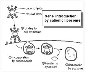
Cationic lipid transfection reagents are suitable for transfecting into a wide variety of dividing cell cultures. Commercial examples include: Lipofectamine / L2000, Dharmafect, iFect, and TransIT TKO. Cationic lipids work by forming lipsomal vesicles that house the siRNA payload and bleb their way through the living cell membrane and into the cytoplasm. The efficiency of this process must be determined in order to have confidence in the knockdown effects. There are numerous commercial sources for transfection reagents for good reason; there are numerous cell types and lipsome structure will influence transfection efficiency in the multitude of experimental cell types that exist.

  • Polymeric

Polymeric formulations have been developed and optimized for transfection of shRNA plasmid DNA into the nucleus of cultured eukaryotic cells by vendors such as Open Biosystems. Cationic lipids but not polyethylenimine or polylysine prevent transgene expression when complexes are injected in the nucleus (Pollard et al 1998). Polymers but not cationic lipids promote gene delivery from the cytoplasm to the nucleus and transgene expression in the nucleus is prevented by complexation with cationic lipids but not with cationic polymers.

shRNA Vector Transfection

  • In a six well tissue culture plate, grow cells to a 50-70% confluency in antibiotic-free normal growth medium supplemented with FBS.

NOTE: This protocol is recommended for a well from a 6 well tissue culture plate. Adjust cell and reagent amounts proportionately for wells or dishes of different sizes.

NOTE: Healthy and subconfluent cells are required for successful transfection experiments. It is recommended to ensure cell viability one day prior to transfection.

Prepare the following solutions:

NOTE: The optimal shRNA Plasmid DNA:shRNA Plasmid Transfection Reagent ratio should be determined experimentally beginning with 1 μg of shRNA Plasmid DNA and between 1.0 and 6.0 μl of shRNA Plasmid Transfection Reagent as outlined below. Once the optimal shRNA Plasmid DNA:shRNA Plasmid Transfection Reagent ratio has been identified for a given cell type, the appropriate amount of shRNA Plasmid DNA/shRNA Plasmid Transfection Reagent complex used per well should be tested to determine which amount provides the highest level of transfection efficiency. For example, if the optimal shRNA Plasmid DNA:shRNA Plasmid Transfection Reagent ratio is 1 μg:1 μl, then amounts ranging from 0.5 μg/0.5 μl to 2.0 μg/2.0 μl should be tested.

Solution A: For each transfection, dilute 10 μl of resuspended shRNA Plasmid DNA (i.e. 1 μg shRNA Plasmid DNA) into 90 μl shRNA Plasmid Transfection Medium (serum antibiotic free medium).

Solution B: For each transfection, dilute 1 – 6 μl of shRNA Plasmid Transfection Reagent with enough shRNA Plasmid Transfection Medium to bring final volume to 100 μl.

NOTE: Do not add antibiotics to the shRNA Plasmid Transfection Medium.

NOTE: Optimal results may be achieved by using siliconized microcentrifuge tubes.

NOTE: Although highly efficient in a variety of cell lines, not all shRNA Plasmid Transfection Reagents may be suitable for use with all cell lines.

  • Add the shRNA Plasmid DNA solution (Solution A) directly to the dilute shRNA Plasmid Transfection Reagent (Solution B) using a pipette. Mix gently by pipetting the solution up and down and incubate the mixture 15-45 minutes at room temperature.
  • Wash the cells twice with 2 ml of shRNA Transfection Medium. Aspirate the medium and proceed immediately to the next step. NOTE: Do not use PBS as the residual phosphate may compete with DNA and bind the shRNA Plasmid Transfection Reagent, thereby reducing the transfection efficiency.

NOTE: Do not use PBS as the residual phosphate may compete with DNA and bind the shRNA Plasmid Transfection Reagent, thereby reducing the transfection efficiency. For each transfection, add 0.8 ml shRNA Plasmid Transfection Medium to well.

  • For each transfection, add 0.8 ml shRNA Plasmid Transfection Medium to well.
  • Add the 200 μl shRNA Plasmid DNA/shRNA Plasmid Transfection Reagent Complex (Solution A + Solution B) dropwise to well, covering the entire layer.
  • Gently mix by swirling the plate to ensure that the entire cell layer is immersed in solution.
  • Incubate the cells 5-7 hours at 37° C in a CO2 incubator or under conditions normally used to culture the cells.

NOTE: Longer transfection times may be desirable depending on the cell line.

  • Following incubation, add 1 ml of normal growth medium containing 2 times the normal serum and antibiotics concentration (2x normal growth medium).
  • Incubate the cells for an additional 18-24 hours under conditions normally used to culture the cells.

Aspirate the medium and replace with fresh 1x normal growth medium.

  • Assay the cells using the appropriate protocol 24-72 hours after the addition of fresh medium in the step above.

NOTE: Controls should always be included in shRNA experiments. Control shRNAs are available as 20 μg. Each encode a scrambled shRNA sequence that will not lead to the specific degradation of any known cellular mRNA.

NOTE: For Western blot analysis prepare cell lysate as follows: Wash cells once with PBS. Lyse cells in 300 μl 1x Electrophoresis Sample Buffer (sc-24945) by gently rocking the 6 well plate or by pipetting up and down. Sonicate the lysate on ice if necessary.

NOTE: For RT-PCR analysis isolate RNA using the method described by P. Chomczynski and N. Sacchi (1987. Single-step method of RNA isolation by acid guanidinium thiocyanate-phenol-chloroform extraction. Anal. Biochem. 162: 156-159) or a commercially available RNA isolation kit.

Lentivrus Infection

Materials:

  • Target cells are SH-SY5Y human neuroblastoma cells.
  • Complete medium: DMEM F-12 medium with 10% serum and Penicillin/Streptomycicn
  • Vectors are copGFP control Lentiviral Particles (sc-108084)
  • Use 24 well plate. Each well has 1.9 mm2, good for working with 0.5 ml medium.

Concentration Considerations:

  • Test 5μg/ml Polybrene
  • Each well contains 4×10⁴ cells. Will use Viral Particles at a quantity of 4×10⁵ infection units. The concentration of provided viral particles was 5000 infections units per μl, so I will try 100 μl. The MOI is 10.

Day 1 Seed Cells

  • Plate target cells in 12 well plate 24 hours prior to viral infection. Each well contains 4×10⁴ cells in 0.5 ml complete medium.

Day 2 Lentiviral Infection

  • Monitor the seeded cells and make sure that the cells are around 50% confluent.
  • Bring Polybrene (sc-134220) to room temperature, and complete medium to 37°C.
  • Prepare a mixture of complete medium with Polybrene at a final concentration of 5 μg/ml.
  • Remove media from plate wells and replace with 1ml of Polybrene/media mixture per well.
  • Put the plate back to the incubator until lentiviral particles were thawed.
  • Thaw lentiviral particles at room temperature and mix gently before use. This takes about 5 minutes.
  • Infect cells by adding the indicated amount of shRNA lentiviral particles to the culture.
  • Swirl the plate gently to mix and incubate overnight.

NOTE: Lentiviral particles were thawed at room temperature. As soon as the vial is thawed, Lentiviral particles were immediately added to the plate to avoid prolonged exposure of the particles to ambient temperature. Did not use ice.

Day 3 Change Medium

  • Remove the culture medium and replace with 1 ml of complete medium (without Polybrene).
  • Incubate the cells for 2 days.
  • Examine GFP positive cells under microscope. Found that around 80% cells were GFP-positive, the signal was weak.

Day 5 and on Culturing of Cells before Selection

  • Starting from Day 5, change to fresh medium and wait for cells to reach confluency in order to get enough cells for the selection.

Changed medium on day5, day8.

shRNA Controls

Negative Controls

  • Untreated Cells. Untreated cells will provide a reference point for comparing all other samples.
  • Empty construct, containing no shRNA insert; The empty viral particles or DNA are a useful negative control that will not activate the RNAi pathway because it does not contain an shRNA insert. It will allow for observation of cellular effects of the transduction/transfection process. Cells transduced/transfected with the empty control provide a useful reference point for comparing specific knockdown.
  • Non-targeting shRNA; This non-targeting shRNA is a useful negative control that will activate RISC and the RNAi pathway, but does not target any human or mouse genes. The short hairpin sequence cotnains 5 base pair mismatches to any known human or mouse gene. This allows for examination of the effects of shRNA transduction/transfection on gene expression. Cells transduced/transfected with the non-target shRNA will also provide useful reference for interpretation of knockdown.

Positive Controls

  • Positive shRNA knockdown control; This control contains shRNA sequence that targets GFP expression. This shRNA control has been experimentally shown to reduce GFP expression. This control serves to quickly visualize knockdown in cells expressing GFP.
  • Positive shRNA knockdown control; This control contains shRNA sequence that targets eGFP expression (GenBank Accession # pEGFP U476561). The shRNA has been experimentally shown to reduce eGFP expression by 90% in C166-GFP mouse fibroblast cells 48 hours post-transduction by mRNA transcript level. This control serves to quickly visualize knockdown in cells expressing eGFP.
  • Positive reporter vector or lentiviral particles; This is a useful positive control for measuring transduction/transfection efficiency and optimizing shRNA delivery. The GFP Control contains a gene encoding GFP, driven by the CMV promoter. This control provides fast visual confirmation of successful transduction/transfection.

copGFP

The copGFP protein is a novel natural green monomeric green fluorescent protein cloned from copepod Pontellina plumata, a type of plankton. The copGFP protein is a non-toxic, non-aggregating protein with fast protein maturation, high stability at a wide range of pH (pH 4-12), and fluorescent properties that do not require any additional cofactors or substrates.

Due to its exceptional properties, copGFP is an excellent fluorescent marker that can be used instead of EGFP (the widely used Aequrea victoria GFP mutant) for monitoring delivery of lentiviral constructs into cells. The copGFP protein has a very bright fluorescence that exceeds the brightness of EGFP by approximately a third.

The copGFP protein emits green fluorescence with the following characteristics:

  • emmision wavelength max – 502 nm
  • excitation wavelength max – 482 nm
  • quantum yield – 0.6
  • extinction coefficient – 70,000 M-1 cm-1

When assaying cells, DO NOT fix with methanol and minimize exposure to light. PFA/Formalin fixation works.

Evrogen

Factors Influencing Successful Transfection

Concentration and purity of nucleic acids

Determine the concentration of your DNA using 260 nm absorbance. Avoid cytotoxic effects by using pure preparations of nucleic acids.

DNA:In terms of plasmid preparation, McManus Lab has not observed a need to use E. coli cells that are highly defective for recombination. High DNA quality usually means high transfection efficiency. All DNA preparations should be performed by Cesium prep or endotoxin-free ion exchange plasmid purification methods. If poor transfection is consistently observed, it may be worth performing a additional clean-up of the DNA. The transfection protocols described here are sensitive to the amount of DNA. It is important to optimize DNA:Transfection Reagent ratios.

Transfection in serum-free media

The highest transfection efficiencies can be obtained if the cells are exposed to the transfection complexes in serum free conditions followed by the addition of medium containing twice the amount of normal serum to the complex medium 3–5 hrs post transfection (leaving the complexes on the cells). However, the transfection medium can be replaced with normal growth medium if high toxicity is observed.

No antibiotics in transfection medium

The presence of antibiotics can adversely affect the transfection efficiency and lead to increased toxicity levels in some cell types. It is recommended that these additives be initially excluded until optimized conditions are achieved, then these components can be added, and the cells can be monitored for any changes in the transfection results.

High protein expression levels

Some proteins when expressed at high levels can by cytotoxic; this effect can also be cell line specific.

Cell history, density, and passage number

It is very important to use healthy cells that are regularly passaged and in growth phase. The highest transfection efficiencies are achieved if cells are plated the day before. However, adequate time should be allowed to allow the cells to recover from the passaging (generally >12 hours). Plate cells at a consistent density to minimize experimental variation. If transfection efficiencies are low or reduction occurs over time, thawing a new batch of cells or using cells with a lower passage number may improve the results.

References

  1. Murphy S, Altruda F, Ullu E, Tripodi M, Silengo L, and Melli M. DNA sequences complementary to human 7 SK RNA show structural similarities to the short mobile elements of the mammalian genome. J Mol Biol. 1984 Aug 25;177(4):575-90. DOI:10.1016/0022-2836(84)90038-x | PubMed ID:6548262 | HubMed [Paper1]
  2. Czauderna F, Santel A, Hinz M, Fechtner M, Durieux B, Fisch G, Leenders F, Arnold W, Giese K, Klippel A, and Kaufmann J. Inducible shRNA expression for application in a prostate cancer mouse model. Nucleic Acids Res. 2003 Nov 1;31(21):e127. DOI:10.1093/nar/gng127 | PubMed ID:14576327 | HubMed [Paper2]
  3. Koper-Emde D, Herrmann L, Sandrock B, and Benecke BJ. RNA interference by small hairpin RNAs synthesised under control of the human 7S K RNA promoter. Biol Chem. 2004 Sep;385(9):791-4. DOI:10.1515/BC.2004.103 | PubMed ID:15493873 | HubMed [Paper3]
  4. Whither RNAi?. Nat Cell Biol. 2003 Jun;5(6):489-90. DOI:10.1038/ncb0603-490 | PubMed ID:12776118 | HubMed [Paper4]
  5. Pollard H, Remy JS, Loussouarn G, Demolombe S, Behr JP, and Escande D. Polyethylenimine but not cationic lipids promotes transgene delivery to the nucleus in mammalian cells. J Biol Chem. 1998 Mar 27;273(13):7507-11. DOI:10.1074/jbc.273.13.7507 | PubMed ID:9516451 | HubMed [Paper5]

TissueCulture: splitting cells-PDF

Concept

Tissue culture cells need to be “split” to keep them alive. The process involves taking a portion of the cells and resuspending them in fresh culture media so they can grow. If they’re left too long in the culture media, they will die, so this process needs to be repeated to keep them alive. They’re kept in DMEM, which is red in color. Once it turns orange, the cells are ready to be split again. The DMEM must be used at room temperature or, optimally, at 37°C

Procedure

As per t175 dish

  1. Take the stock plate from the incubator
  2. In the tissue culture hood and being careful not to touch the cells (on the bottom), aspirate off all the media (use a clean tip!)
  3. Add 10 ml of PBS carefully (so as not to kill the cells, use the “gravity” setting on the pipettor)
  4. Add 500 ul of trypsin-EDTA
  5. Swirl around
  6. Put at 37°C for 5 minutes to let the trypsin work
  7. Add 10 ml of DMEM+FBS+glutamine+antibiotics to the trypsinized cells
  8. Add 10 ml of DMEM+FBS+glutamine+antibiotics to a new, labelled petri dish
  9. Add 1 ml of the trypsinized cells to the new dish, discard the rest (aspirate off then throw away dish)
  10. Incubate at 37°C until desired confluency

To add

DMEM needs 50 ml FBS, 5 ml glutamine, 5 ml antibiotics

The final concentration of FBS should be 10% v/v The final concentration of antibiotics should be 1% v/v

Use this media for everything except lipofectamine transfection Low serum media w/o antibiotics, such as Opti-MEM, should be used during transfection, and transduction.

You can use Hela most of the time, but Heck is also good?

TissueCulture: Thawing cells-PDF

Overview

This short protocol describes how to freeze down and thaw (or bring up) mammalian cells (e.g. HeLa, KEK293, CHO, Jurkat T cells).

Materials

List reagents, supplies, and equipment necessary to perform the protocol here.

  • 10-50 ml centrifuge tube
  • Cell culture medium (depends on cell line/cells)
  • Trypsin-EDTA (e.g. from PAA, L11-004) – for adherent cells
  • Basic TC centrifuge (0 – 2500 rpm)

Thawing

  1. Setup two vials of cold (4 C) media (10 ml) in a 15 ml tube
  2. Warm remaining media (~5-8 ml) in a 15 ml tube to 37C
  3. When ready to thaw, remove the vial of cells from the liquid nitrogen
    Keep frozen cells on dry ice until ready for thawing.
  4. Swirl the vial in the water bath (37C) until a small ice chip is left in the tube (~1 min)
  5. Place the vial in the hood and clean it with 70% ethanol
  6. Immediately remove the contents of the vial and place it into the cold media
  7. Rinse the tube down with some of the cold media from the second vial
  8. Spin down cells at 2000 rpm (80-100 x g) for 5 min
  9. Aspirate off the cold media and resuspend the cells in the warm media
  10. Transfer the cells and the media into a petri dish
  11. Count cells
  12. Place in incubator
  13. Change the media once the cells have attached to the plate
    Removes remaining DMSO
  14. Allow cells to grow to 80-90% confluency
  15. Split cells (1:3) into Petri dishes
  16. Continue to split cells and freeze down as needed

Notes

  1. List troubleshooting tips here.
  2. You can also link to FAQs/tips provided by other sources such as the manufacturer or other websites.
  3. Anecdotal observations that might be of use to others can also be posted here.

Alternative Thawing

  • From step 5 above, using a 1 ml pipet add 1 ml of the cold media from the 15 ml tube drop by drop to the vial (about 1 drop every 10 seconds).
  • Plate immediately with the cold media and change the media once cells have attached (12 – 24 hours later) to remove the DMSO that was in the freezing media. Usually, a 1:3 ratio is good for MC3T3s and a 1:4 ratio for MLOY4s.

Hop DNA Isolation-PDF

Hop DNA Extraction Protocol

  1. Obtain an adequate amount (~ 1g) of fresh hop leaves and crush them with liquid Nitrogen and a small amount of Carborundum powder (fine 320 grit).
  2. Assume 90% of mass is water weight.
  3. Add 3.3 ml of buffer per gram of wet (16 ml per gram of dried) hop leaves, and incubate for 1 to 4 hours at 60-65°C.
  4. Transfer 900 μl into fresh tube
  5. add 600 μl of 24:1 CHCl3:octanol and invert gently (do NOT vortex!).
  6. Centrifuge at 5000g for 10 minutes.
  7. Transfer supernatant (800 μl) into new 2-ml tube.
  8. Add 5μl of RNAase and incubate at 37°C for 30 minutes (or more).
  9. Add 0.6 volumes Isopropanol and mix gently by inverting the tubes. Check for DNA precipitation.
  10. Spin down for 10 min. at RT.
  11. Add 500 μl wash buffer and incubate 10 min. at RT.
  12. Carefully remove wash buffer. Don’t lose DNA pellet!
  13. Briefly centrifuge to collect pellet at bottom of tube – remove any remaining wash buffer.
  14. Dry pellet at RT or 50°C to speed up.
  15. Add 100 μl ddH2O to dissolve DNA.
  16. Store at -20°C until needed.
  17. Run electrophoresis for analysis.

Prepared solutions

  1. Buffer: 100 ml: 50 mM Tris/HCl (ph 8.0), 1.8 M NaCl, 50 mM EDTA. Then add 10 mg/ml of CTAB ( 200 mg per 20 ml buffer, final conc. = 1%) and 1 μl/ml 2-mercaptoethanol (20 μl to 20 ml buffer; final conc. = 0.1%).
  2. Wash buffer 100 ml: 200 μl 5M NH4OAc (final conc. = 10 mM), 76.0 ml abs. ethanol (final conc. = 76%), and 23.8 ml of sterilized water.

Transformation Protocol for Tobacco-PDF

Plant material:In vitro plants of Nicotiana tabacum L. cv Wisconsin 38

Day 1 Prepare bacteria: • Streak YM plus antibiotic plates with bacteria. • Incubate plates for 2-3 days at 28°C.

Day 3 Transformation:

• Measure about 20mL Minimal A medium for each bacterial strain.

• Scrape or wash bacteria from plate with sterile loop and suspend in 20mL of Minimal A.

• Adjust density to an OD600 0.9-1.0.

• Take first healthy fully expanded leaves from 4-5 week old tissue culture grown tobacco plant, cut into 0.5cm squares (or can use a cork borer, which is about 1.0cm diameter) in deep petri dish, under sterile RMOP liquid medium. Store tissue pieces in RMOP in deep petri dish.

• Transfer leaf pieces (about 20 per transformation) to deep petri dish containing bacterial suspension.

• Swirl to ensure bacteria has contacted cut edge of leaf and let stand for 5 minutes.

• Remove leaf pieces from suspension and blot dry on filter paper or on the edge of the container.

• Place leaf pieces with adaxial side (upper leaf surface) on solid RMOP, about 10 pieces per plate.

• Incubate plates in dark at 28°C for: 2-3 days A. tumefaciens 5 days S. meliloti 5 days M. loti 5-11 days Rhizobium sp. NGR234

Day 6-14 Selection:

• Transfer leaf pieces onto solid RMOP-TCH, with abaxial surface (lower surface of leaf) in contact with media.

• Incubate plates for 2-3 weeks in the light at 28°C, with 16 hours daylight per day.

• Subculture every 2 weeks.

Plantlet formation:

• When shoots appear transfer to MST-TCH pots.

• Incubate plantlets with 16 hours daylight for 1-2 weeks.

• When roots form the plants can be transferred to soil in the glasshouse.

• Plants can be maintained in tissue culture but will need to be subcultured every two weeks. This is done by cutting the growing tip of plant, removing excess leaves and transferring this to fresh media.


Media and Solutions for Tobacco Transformation

YM Media

Mannitol 10g

Yeast extract 0.4g

K2HPO4 (10% w/v stock) 1 ml

KH2PO4 (10% w/v stock) 4 ml

NaCl (10% w/v stock) 1 ml

MgSO4.7H2O (10% w/v stock) 2 ml

• Add Water to 1L

• pH 6.8

• Agar 15g/L

• Autoclave

• When ready to pour add antibiotic selection if required

Keep poured plates for 2 days at room temperature to visualise any contamination, then store at 4°C.


RMOP + RMOP-TCH media (Svab, et al, 1975)

Sucrose 30g 3%

Myo-inositol 100mg 0.1%

MS Macro 10x 100mL 1x

MS Micro 1000x 1mL 1x

Fe2EDTA Iron 100x 10mL 1x

Thiamine-HCl (10mg/mL stock) 100μL 1mg

NAA (1mg/mL stock) 100μL 0.1mg

BAP (1mg/mL stock) 1mL 1mg

• pH 5.8

• Phytagel 2.5g/L for solid

• autoclave

• for RMOP-TCH, when ready to pour add:

• Timentin (200mg/mL stock) 1mL

• Claforan (250mg/mL stock) 1mL

• Hygromycin (50mg/mL stock) 1mL


BAP (1mg/ml) (6-Benzylaminopurine) Add 1N KOH drop wise to 100mg BAP until dissolved. Make up to 100mL

with Milli-Q H2O

• Store 4°C

NAA (1mg/ml) (Naphthalene acetic acid)

Dissolve 100mg NAA in 1mL absolute ethanol. Add 3mL 1N KOH. Make up

to 80mL with Milli-Q H2O. Adjust pH to 6.0 with 1N HCl, make up to 100mL

with Milli-Q H2O.

• Store 4°C

Cefotaxamine® (250mg/ml)

Add 8ml sterile Milli-Q H2O to 2g Claforan • Store 4°C in dark

Timentin® (200mg/ml)

Add 15ml sterile Milli-Q H2O to 3g Timentin

• Store 4°C

MST + MST-TCH media (Svab, et al, 1975)

Sucrose 30g 3%

MS Macro 10x 100mL 1x

MS Micro 1000x 1mL 1x

Fe2EDTA Iron 100x 10mL 1x

• pH 5.8

• Phytagel 2.5g/L

• autoclave

• for MST-TCH, when ready to pour add:

• Timentin® (200mg/mL stock) 1mL

• Cefotaxamine® (250mg/mL stock) 1mL

• Hygromycin (50mg/mL stock) 1mL

MS Macro 10x (Murashige and Skoog, 1962)


Final concentration 
   10x (g/L)    1 x mM 

KNO3 19.0 18.8

NH4 N03 16.5 20.6

CaCl2.2H2O 4.4 3.0

MgS04.7H2O 3.7 1.5

KH2PO4 1.7 1.25

• Store 4°C

• Substituting chemicals:

• CaCl2 3.3g/L

• MgS04 1.8g/L

MS Micro 1000x (Murashige and Skoog, 1962)

                                                                              Final concentration 
   1000x (g/L)                         1 x μM 

MnS04.4H20 22.3 100

ZnS04.7H20 8.6 30

H3BO3 6.2 100

KI 0.83 5.0

Na2MoO4.2H2O 0.25 1.0

CuSO4.5H2O 25mg 0.1

CoCl2.6H2O 25mg 0.1

• Store 4°C

• Substituting chemicals:

• MnS04.H20 16.9/L

FeSO4EDTA Iron 100x

   g/1L                               1 x mM 

FeS04.7H20 2.78 0.1

Na2EDTA 3.72 0.1

• Store 4°C in dark bottle


References:

Murashige, T. and F. Skoog. 1962. A revised medium for rapid growth and bio assays with tobacco tissue cultures. Phys. Plant. 15: 473-497.

Svab, Z., P. Hajdukiewicz and P. Maliga. 1975. Transgenic tobacco plants by co- cultivation of leaf disks with pPZP Agrobacterium binary vectors. In “Methods in Plant Molecular Biology-A Laboratory Manual”, P. Maliga, D. Klessig, A.. Cashmore, W. Gruissem and J. Varner, eds. Cold Spring Harbor Press: 55- 77.